STAT3 knockdown reduces pancreatic cancer cell invasiveness and matrix metalloproteinase-7 expression in nude mice
- PMID: 21991388
- PMCID: PMC3185063
- DOI: 10.1371/journal.pone.0025941
STAT3 knockdown reduces pancreatic cancer cell invasiveness and matrix metalloproteinase-7 expression in nude mice
Abstract
Aims: Transducer and activator of transcription-3 (STAT3) plays an important role in tumor cell invasion and metastasis. The aim of the present study was to investigate the effects of STAT3 knockdown in nude mouse xenografts of pancreatic cancer cells and underlying gene expression.
Methods: A STAT3 shRNA lentiviral vector was constructed and infected into SW1990 cells. qRT-PCR and western immunoblot were performed to detect gene expression. Nude mouse xenograft assays were used to assess changes in phenotypes of these stable cells in vivo. HE staining was utilized to evaluate tumor cell invasion and immunohistochemistry was performed to analyze gene expression.
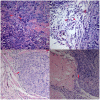
Results: STAT3 shRNA successfully silenced expression of STAT3 mRNA and protein in SW1990 cells compared to control cells. Growth rate of the STAT3-silenced tumor cells in nude mice was significantly reduced compared to in the control vector tumors and parental cells-generated tumors. Tumor invasion into the vessel and muscle were also suppressed in the STAT3-silenced tumors compared to controls. Collagen IV expression was complete and continuous surrounding the tumors of STAT3-silenced SW1990 cells, whereas collagen IV expression was incomplete and discontinuous surrounding the control tumors. Moreover, microvessel density was significantly lower in STAT3-silenced tumors than parental or control tumors of SW1990 cells. In addition, MMP-7 expression was reduced in STAT3-silenced tumors compared to parental SW1990 xenografts and controls. In contrast, expression of IL-1β and IgT7α was not altered.
Conclusion: These data clearly demonstrate that STAT3 plays an important role in regulation of tumor growth, invasion, and angiogenesis, which could be act by reducing MMP-7 expression in pancreatic cancer cells.
Conflict of interest statement
Figures


Similar articles
-
STAT3-targeting RNA interference inhibits pancreatic cancer angiogenesis in vitro and in vivo.Int J Oncol. 2011 Jun;38(6):1637-44. doi: 10.3892/ijo.2011.1000. Epub 2011 Apr 8. Int J Oncol. 2011. PMID: 21479358
-
[Effect of RNAi-mediated STAT3 gene inhibition on metastasis of human pancreatic cancer cells].Zhonghua Wai Ke Za Zhi. 2008 Jul 1;46(13):1010-3. Zhonghua Wai Ke Za Zhi. 2008. PMID: 19035205 Chinese.
-
Down-regulation of STAT3 expression by vector-based small interfering RNA inhibits pancreatic cancer growth.World J Gastroenterol. 2011 Jul 7;17(25):2992-3001. doi: 10.3748/wjg.v17.i25.2992. World J Gastroenterol. 2011. PMID: 21799645 Free PMC article.
-
Lentivirus-mediated shRNA interference targeting STAT3 inhibits human pancreatic cancer cell invasion.World J Gastroenterol. 2009 Aug 14;15(30):3757-66. doi: 10.3748/wjg.15.3757. World J Gastroenterol. 2009. PMID: 19673016 Free PMC article.
-
MMP-7 marks severe pancreatic cancer and alters tumor cell signaling by proteolytic release of ectodomains.Biochem Soc Trans. 2022 Apr 29;50(2):839-851. doi: 10.1042/BST20210640. Biochem Soc Trans. 2022. PMID: 35343563 Free PMC article. Review.
Cited by
-
Shifting paradigm of developing biologics for the treatment of pancreatic adenocarcinoma.J Gastrointest Oncol. 2017 Jun;8(3):441-448. doi: 10.21037/jgo.2016.10.02. J Gastrointest Oncol. 2017. PMID: 28736631 Free PMC article. Review.
-
Role of STAT3 in cancer metastasis and translational advances.Biomed Res Int. 2013;2013:421821. doi: 10.1155/2013/421821. Epub 2013 Oct 2. Biomed Res Int. 2013. PMID: 24199193 Free PMC article. Review.
-
The Multifaced Role of STAT3 in Cancer and Its Implication for Anticancer Therapy.Int J Mol Sci. 2021 Jan 9;22(2):603. doi: 10.3390/ijms22020603. Int J Mol Sci. 2021. PMID: 33435349 Free PMC article. Review.
-
STAT3 in Cancer-Friend or Foe?Cancers (Basel). 2014 Jul 3;6(3):1408-40. doi: 10.3390/cancers6031408. Cancers (Basel). 2014. PMID: 24995504 Free PMC article.
-
Exocrine pancreatic carcinogenesis and autotaxin expression.PLoS One. 2012;7(8):e43209. doi: 10.1371/journal.pone.0043209. Epub 2012 Aug 29. PLoS One. 2012. PMID: 22952646 Free PMC article.
References
-
- Jemal A, Siegel R, Xu J, Ward E. Cancer statistics, 2010. CA Cancer J Clin. 2010;60:277–300. - PubMed
-
- Bowman T, Garcia R, Turkson J, Jove R. STATs in oncogenesis. Oncogene. 2000;19:2474–2488. - PubMed
-
- Li WC, Ye SL, Sun RX, Liu YK, Tang ZY, et al. Inhibition of growth and metastasis of human hepatocellular carcinoma by antisense oligonucleotide targeting signal transducer and activator of transcription 3. Clin Cancer Res. 2006;12:7140–7148. - PubMed
-
- Xie TX, Wei D, Liu M, Gao AC, Ali-Osman F, et al. Stat3 activation regulates the expression of matrix metalloproteinase-2 and tumor invasion and metastasis. Oncogene. 2004;23:3550–3560. - PubMed
Publication types
MeSH terms
Substances
LinkOut - more resources
Full Text Sources
Other Literature Sources
Medical
Miscellaneous

