. 2011 Oct;5(10):e1345.
doi: 10.1371/journal.pntd.0001345. Epub 2011 Oct 4.

Human cellular immune response to the saliva of Phlebotomus papatasi is mediated by IL-10-producing CD8+ T cells and Th1-polarized CD4+ lymphocytes

Affiliations

Human cellular immune response to the saliva of Phlebotomus papatasi is mediated by IL-10-producing CD8+ T cells and Th1-polarized CD4+ lymphocytes

Maha Abdeladhim et al. PLoS Negl Trop Dis. 2011 Oct.

Abstract

Background: The saliva of sand flies strongly enhances the infectivity of Leishmania in mice. Additionally, pre-exposure to saliva can protect mice from disease progression probably through the induction of a cellular immune response.

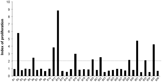
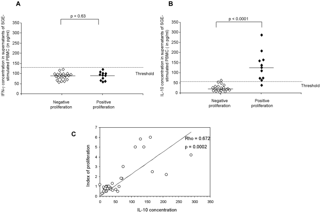
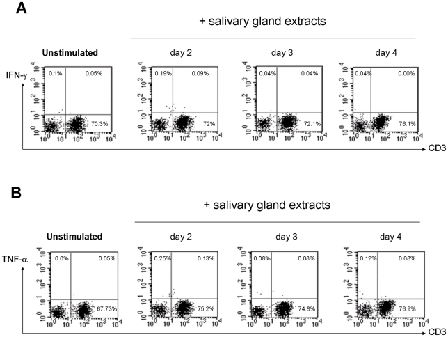
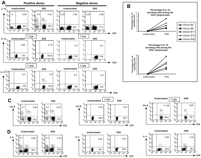
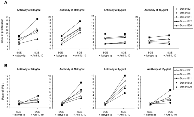
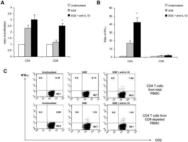
Methodology/principal findings: We analysed the cellular immune response against the saliva of Phlebotomus papatasi in humans and defined the phenotypic characteristics and cytokine production pattern of specific lymphocytes by flow cytometry. Additionally, proliferation and IFN-γ production of activated cells were analysed in magnetically separated CD4+ and CD8+ T cells. A proliferative response of peripheral blood mononuclear cells against the saliva of Phlebotomus papatasi was demonstrated in nearly 30% of naturally exposed individuals. Salivary extracts did not induce any secretion of IFN-γ but triggered the production of IL-10 primarily by CD8+ lymphocytes. In magnetically separated lymphocytes, the saliva induced the proliferation of both CD4+ and CD8+ T cells which was further enhanced after IL-10 blockage. Interestingly, when activated CD4+ lymphocytes were separated from CD8+ cells, they produced high amounts of IFN-γ.

Conclusion: Herein, we demonstrated that the overall effect of Phlebotomus papatasi saliva was dominated by the activation of IL-10-producing CD8+ cells suggesting a possible detrimental effect of pre-exposure to saliva on human leishmaniasis outcome. However, the activation of Th1 lymphocytes by the saliva provides the rationale to better define the nature of the salivary antigens that could be used for vaccine development.


Conflict of interest statement

The authors have declared that no competing interests exist.

Figures

Figure 1
Figure 1. Salivary gland extracts of Phlebotomus papatasi induce proliferation of PBMC in previously exposed individuals.
Peripheral blood mononuclear cells (PBMC) were isolated from 36 volunteer donors; 10 donors (B1 to B10) were from a non endemic region for ZCL and 26 individuals (B11 to B36) were from endemic areas. PBMC (0.5×10⁶ cells/mL) were stimulated in 96-well plates with salivary gland extracts at one gland/ml during five days. Proliferative responses were assessed by (3H) thymidine uptake. Results were expressed as an index of proliferation (mean counts of triplicates in antigen-stimulated cultures /mean counts of triplicates in unstimulated cultures) in all tested individuals. We designated as positive SGE proliferation the cases exhibiting an index of PBMC proliferation above 2. The indices of proliferation obtained in such donors were relatively low with a median of 3.2 but significantly higher than those obtained in donors with negative SGE responses (p<0.0001).
Figure 2
Figure 2. Salivary gland extracts of Phlebotomus papatasi induce IL-10 secretion by human PBMC in immune individuals.
Purified peripheral blood mononuclear cells (0.5×10⁶ cells/mL) were stimulated with salivary gland extract of P. papatasi. IFN-γ (A) and IL-10 (B) secretion was evaluated in supernatants of cultures by an ELISA test at day 3 and day 2, respectively. Results were expressed as cytokine concentrations in stimulated cultures for individuals with or without PBMC proliferation against salivary gland extract. The threshold was calculated as the 95th percentile of the values obtained in individuals with negative proliferation. Horizontal bars indicate median levels of cytokines. The correlation between IL-10 concentrations in supernatants of stimulated cells and proliferative responses in all tested donors was represented in (C).
Figure 3
Figure 3. Salivary gland extracts of Phlebotomus papatasi did not induce IFN-γ or TNF-α production in stimulated PBMC.
Freshly purified peripheral blood mononuclear cells (0.5×10⁶ cells/mL) from 5 individuals (B2, B6, B11, B12 and B20) were stimulated with salivary gland extracts of P. papatasi at 1 gland/ml during 96h. Intracytoplamic expression of IFN-γ (A) and TNF-α (B) was studied from days 2 through 4 by flow cytometry in CD3 membrane stained-cells. The result from individual B11 is shown.
Figure 4
Figure 4. Salivary gland extracts of Phlebotomus papatasi activate IL-10- and IL-4-producing CD8 T lymphocytes.
Freshly purified peripheral blood mononuclear cells (PBMC at 0.5×10⁶ cells/mL) from 4 (B2, B6, B11 and B12) or 5 individuals (B2, B6, B11, B12 and B20) were stimulated with salivary gland extract of P. papatasi at 1 gland/ml. Intracytoplamic expression of IL-10 (A and B), Granzyme B (C) and IL-4 (D) was studied by flow cytometry at day 3 in CD3, CD4 and CD8 membrane stained-cells. Cytokine staining was analyzed in the lymphocyte gate, on CD4+CD3+ gated cells (T CD4) or on CD8+CD3+ gated cells (T CD8). In panel A, C and D, the results from one representative donor (B11) who exhibits proliferative response to SGE are shown. Additionally, the result from the donor A3 with no proliferative response to SGE, is shown in panel A.
Figure 5
Figure 5. Effect of anti-IL-10 blocking antibody on lymphocytes stimulated with salivary gland extracts of Phlebotomus papatasi.
Total PBMC (0.5×10⁶ cells/mL) from donors B2, B6, B11, B12 and B20 were stimulated with salivary gland extract (1 gland/ml) for 5 days in the presence of blocking anti-IL-10 or isotype control at the indicated concentrations. (A) Proliferative responses were assessed by (3H) thymidine uptake. Results were expressed as index of proliferation: mean counts of triplicates in antigen-stimulated cultures /mean counts of triplicates in unstimulated cultures. (B) IFN-γ secretion was evaluated in supernatants of cultures at day 3 using an ELISA test. Results were expressed as a ratio of cytokine levels in stimulated/unstimulated cultures.
Figure 6
Figure 6. Effect of IL-10 blockage and CD8 cell depletion on lymphocytes stimulated with salivary gland extracts of Phlebotomus papatasi.
Total peripheral blood mononuclear cells (PBMC) or PBMC depleted of CD4+ or CD8+ T lymphocytes from donors B2, B6, B11, B12 and B20 (0.5×10⁶ cells/mL) were stimulated with salivary gland extract (1 gland/ml) for 5 days in the presence or not of anti-IL-10 blocking antibody at 500ng/ml. (A) Proliferative responses were assessed by [3H] thymidine uptake. Results were expressed as index of proliferation: mean counts of triplicates in antigen-stimulated cultures /mean counts of triplicates in unstimulated cultures. (B) IFN-γ secretion was evaluated in supernatants of cultures at day 3 using an ELISA test. Results were expressed as a ratio of cytokine levels in stimulated/unstimulated cultures. * p<0.05 when compared to the condition with SGE. Data are representative of 5 distinct experiments. (C) Intracytoplamic expression of IFN-γ was studied by flow cytometry at day 3 in total (upper panels) or CD8-depleted PBMC (lower panels). Cytokine staining was analyzed in the lymphocyte gate, on CD4+CD3+ gated cells. Result from one representative individual (B11) is shown.

References

    1. Herwaldt BL. Leishmaniasis. Lancet. 1999;354:1191–1199. - PubMed
    1. Ribeiro JM. Role of saliva in blood-feeding by arthropods. Annu Rev Entomol. 1987;32:463–478. - PubMed
    1. Charlab R, Valenzuela JG, Rowton ED, Ribeiro JM. Toward an understanding of the biochemical and pharmacological complexity of the saliva of a hematophagous sand fly Lutzomyia longipalpis. Proc Natl Acad Sci U S A. 1999;96:15155–15160. - PMC - PubMed
    1. Sacks D, Kamhawi S. Molecular aspects of parasite-vector and vector-host interactions in leishmaniasis. Annu Rev Microbiol. 2001;55:453–483. - PubMed
    1. Titus RG, Ribeiro JM. Salivary gland lysates from the sand fly Lutzomyia longipalpis enhance Leishmania infectivity. Science. 1988;239:1306–1308. - PubMed

Publication types