Comparative Study
. 2011;13(5):R164.
doi: 10.1186/ar3483. Epub 2011 Oct 13.

Bone destruction by receptor activator of nuclear factor κB ligand-expressing T cells in chronic gouty arthritis

Affiliations
Comparative Study

Bone destruction by receptor activator of nuclear factor κB ligand-expressing T cells in chronic gouty arthritis

Sung-Ji Lee et al. Arthritis Res Ther. 2011.

Abstract

Introduction: The purpose of this study was to analyze the cellular expressions of pro-resorptive cytokines in gouty tophus tissues, to determine the capacity of monosodium urate monohydrate (MSU) crystals to induce these cytokines, and to understand the mechanisms of bone destruction in chronic gout.

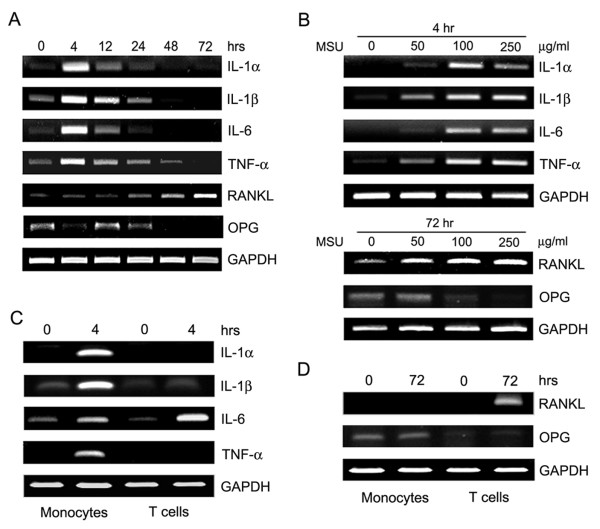
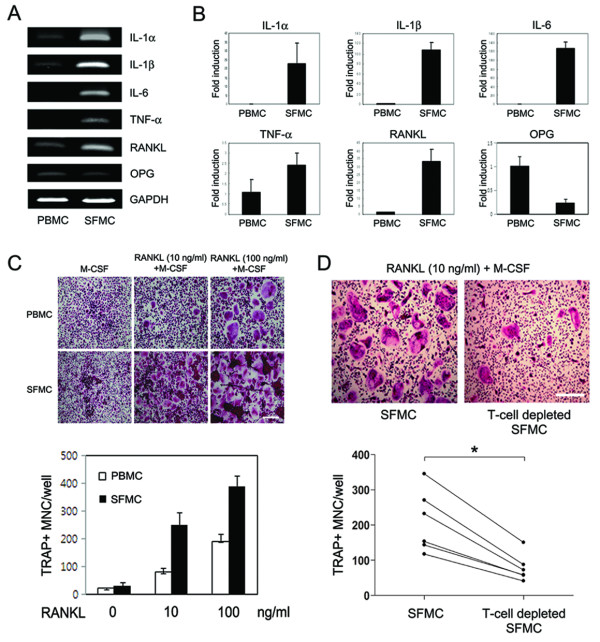
Methods: Fourteen fixed, paraffin-embedded, uninfected tophus samples were analyzed immunohistochemically. Peripheral blood mononuclear cells (PBMCs) were cultured in vitro with MSU crystals, and gene expression was assessed by reverse transcription-polymerase chain reaction. In vitro osteoclastogenesis was performed using PBMCs and synovial fluid mononuclear cells (SFMCs).

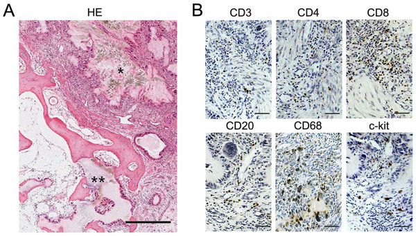
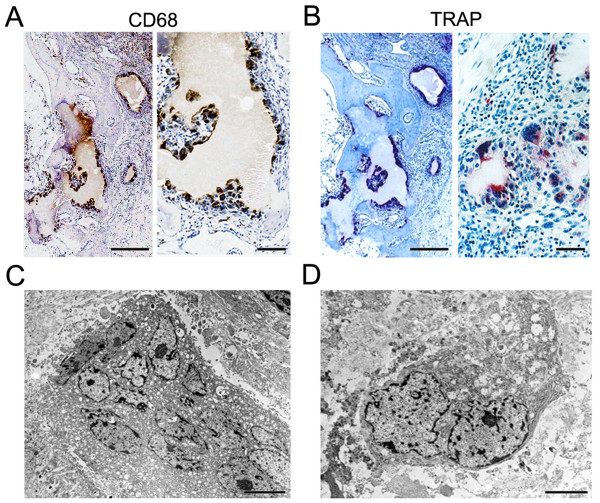
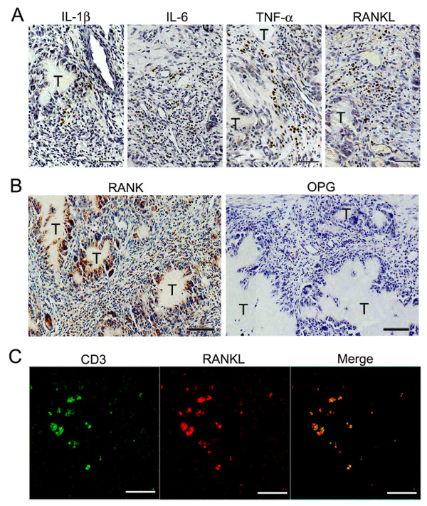
Results: CD4+ T cells, CD8+ T cells, CD20+ B cells and mast cells infiltrated tophus tissues. Tartrate-resistant acid phosphatase (TRAP)+ osteoclasts were present around tophi and in osteolytic lesions. Interleukin (IL)-1, IL-6 and tumor necrosis factor (TNF)-alpha were produced from infiltrated mononuclear cells, whereas receptor activator of nuclear factor κB ligand (RANKL) was strongly expressed in T cells. However, osteoprotegerin (OPG) was not or was weakly expressed in tophus tissues. MSU crystals induced the expressions of IL-1, IL-6, TNF-alpha and RANKL in PBMCs, but inhibited OPG expression. In addition, the pro-resorptive cytokines were highly expressed in SFMCs of gouty arthritis patients. Furthermore, in vitro osteoclastogenesis was enhanced in SFMC cultures, but inhibited in T cell-depleted SFMC cultures.

Conclusions: Our study demonstrates that RANKL-expressing T cells and TRAP+ osteoclasts are present within gouty tophus tissues, and that infiltrating cells express pro-resorptive cytokines. Furthermore, our data show that MSU crystals have the potential to induce pro-resorptive cytokines, and T cells are involved in osteoclastogenesis in chronic gout.


Figures

Figure 1
Figure 1
Infiltration of chronic inflammatory cells in gouty tophus tissues. (a) Tophus tissue sections from patients with chronic gouty arthritis were processed for hematoxylin-eosin (HE) staining. * indicates monosodium urate monohydrate (MSU) crystals, and ** represents osteolytic lesions. (b) Serial sections were processed for immunohistochemical staining using anti-CD3, anti-CD4, anti-CD8, anti-CD20, anti-CD68, and anti-c-kit antibodies (brown). Scale bar in a = 500 μm; scale bars in b = 50 μm.
Figure 2
Figure 2
Localization of CD68+ and tartrate-resistant acid phosphatase (TRAP)+ cells in gouty tophus tissues. (a) Tophus tissue sections from patients with chronic gouty arthritis were processed for immunohistochemical staining using anti-CD68 antibody (brown). (b) Serial sections were stained for TRAP (red). (c, d) Electron microscopic findings of CD68+ cells. Scale bars in left panels of a and b = 500 μm; scale bars in right panels of a and b = 50 μm; scale bars in c and d = 5 μm.
Figure 3
Figure 3
Expression of pro-resorptive cytokines in gouty tophus tissues. (a, b) Tophus tissue sections from patients with chronic gouty arthritis were processed for immunohistochemical staining using anti-interleukin (IL)-1β, anti-IL-6, anti-tumor necrosis factor (TNF)-α, anti-receptor activator of nuclear factor κB (RANK), anti-osteoprotegerin (OPG), and anti-receptor activator of nuclear factor κB ligand (RANKL) antibodies (brown). T, gouty tophus. (c) Immunofluorescent stains for CD3 (green) and RANKL (red) in tophaceous gout tissues. Scale bars in a, b, and c = 50 μm.
Figure 4
Figure 4
Expression of pro-resorptive cytokines by monosodium urate monohydrate (MSU) crystals. (a) Freshly isolated peripheral blood mononuclear cells (PBMCs) from healthy control subjects were cultured for the indicated times (hours) in the presence of MSU crystals (100 μg/ml). (b) PBMCs from same donors were cultured for the indicated times (that is, 4 hours for cytokines and 72 hours for receptor activator of nuclear factor κB ligand RANKL) at various MSU crystal concentration (μg/ml). (c, d) Monocytes and T cells were isolated from PBMCs by magnetic-activated cell sorting. Freshly isolated monocytes and T cells from healthy control subjects were cultured for the indicated times (hours) in the presence of MSU crystals (100 μg/ml). Total RNA was collected at each time point and at the different MSU crystal concentrations. Reverse transcription-polymerase chain reaction (RT-PCR) was performed to determine the expressions of interleukin (IL)-1α, IL-β, IL-6, tumor necrosis factor (TNF)-α, osteoprotegerin (OPG), and RANKL. Results are representative of three independent experiments.
Figure 5
Figure 5
Pro-resorptive cytokine expression and osteoclastogenesis in the synovial fluid of patients with gouty arthritis. Six paired samples of peripheral blood and synovial fluid were obtained from gouty arthritis patients with knee effusion. (a, b) Expression of mRNA for interleukin (IL)-1α, IL-β, IL-6, tumor necrosis factor (TNF)-α, receptor activator of nuclear factor κB ligand (RANKL) and osteoprotegerin (OPG) in peripheral blood mononuclear cells (PBMCs) and synovial fluid mononuclear cells (SFMCs). Reverse transcription-polymerase chain reaction (RT-PCR) (a) and real-time PCR (b) were performed to determine the mRNA expression of each gene. (c, d) T cell involvement in osteoclastogenesis of SFMCs from patients with gouty arthritis. PBMCs, SFMCs, and T cell-depleted SFMCs (3 × 10⁵ cells/well) were cultured for 10 days in the presence of macrophage colony-stimulating factor (M-CSF; 30 ng/ml) at the different RANKL concentrations and then were stained for tartrate-resistant acid phosphatase (TRAP). TRAP-positive multinucleated cells (MNCs) were counted in triplicate. Results are representative of six independent experiments (a and upper panels in c and d). Data in b and lower panel in c are means ± SEMs. Scale bars in c and d = 50 μm. * = P < 0.05, by Wilcoxon signed rank test.

Comment in

Similar articles

Cited by

References

    1. Richette P, Bardin T. Gout. Lancet. pp. 318–328. - PubMed
    1. Rider TG, Jordan KM. The modern management of gout. Rheumatology (Oxford) pp. 5–14. - PubMed
    1. Nakayama DA, Barthelemy C, Carrera G, Lightfoot RW Jr, Wortmann RL. Tophaceous gout: a clinical and radiographic assessment. Arthritis Rheum. 1984;27:468–471. doi: 10.1002/art.1780270417. - DOI - PubMed
    1. Dalbeth N, Collis J, Gregory K, Clark B, Robinson E, McQueen FM. Tophaceous joint disease strongly predicts hand function in patients with gout. Rheumatology (Oxford) 2007;46:1804–1807. doi: 10.1093/rheumatology/kem246. - DOI - PubMed
    1. Palmer DG, Highton J, Hessian PA. Development of the gout tophus. An hypothesis. Am J Clin Pathol. 1989;91:190–195. - PubMed

Publication types

MeSH terms