

Automatic segmentation of the facial nerve and chorda tympani in pediatric CT scans
- PMID: 21992377
- PMCID: PMC3208411
- DOI: 10.1118/1.3634048
Automatic segmentation of the facial nerve and chorda tympani in pediatric CT scans
Abstract
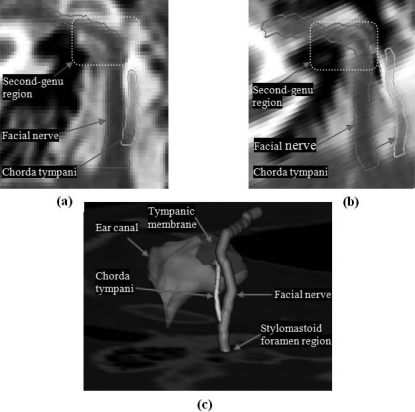
Purpose: Cochlear implant surgery is used to implant an electrode array in the cochlea to treat hearing loss. The authors recently introduced a minimally invasive image-guided technique termed percutaneous cochlear implantation. This approach achieves access to the cochlea by drilling a single linear channel from the outer skull into the cochlea via the facial recess, a region bounded by the facial nerve and chorda tympani. To exploit existing methods for computing automatically safe drilling trajectories, the facial nerve and chorda tympani need to be segmented. The goal of this work is to automatically segment the facial nerve and chorda tympani in pediatric CT scans.
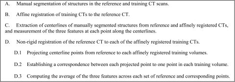
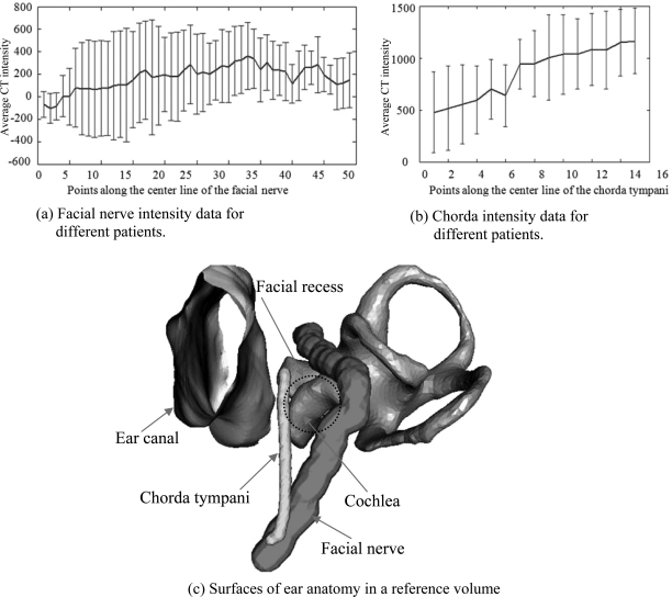
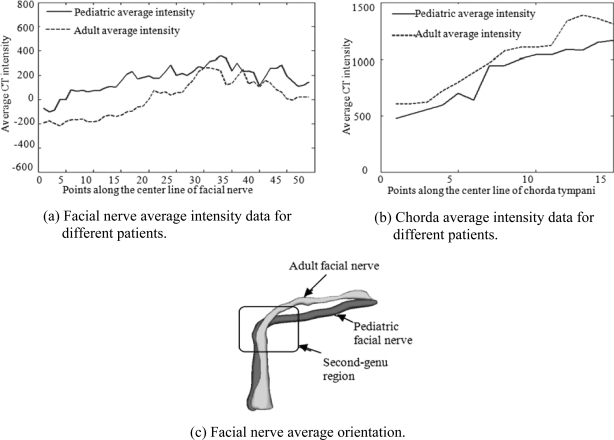
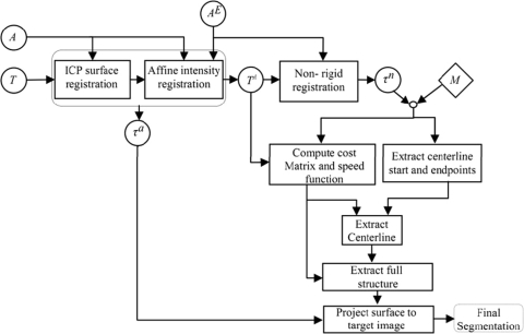
Methods: The authors have proposed an automatic technique to achieve the segmentation task in adult patients that relies on statistical models of the structures. These models contain intensity and shape information along the central axes of both structures. In this work, the authors attempted to use the same method to segment the structures in pediatric scans. However, the authors learned that substantial differences exist between the anatomy of children and that of adults, which led to poor segmentation results when an adult model is used to segment a pediatric volume. Therefore, the authors built a new model for pediatric cases and used it to segment pediatric scans. Once this new model was built, the authors employed the same segmentation method used for adults with algorithm parameters that were optimized for pediatric anatomy.
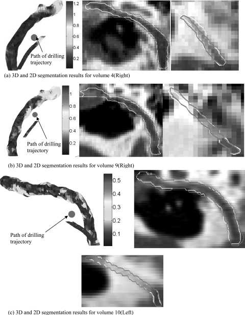
Results: A validation experiment was conducted on 10 CT scans in which manually segmented structures were compared to automatically segmented structures. The mean, standard deviation, median, and maximum segmentation errors were 0.23, 0.17, 0.18, and 1.27 mm, respectively.
Conclusions: The results indicate that accurate segmentation of the facial nerve and chorda tympani in pediatric scans is achievable, thus suggesting that safe drilling trajectories can also be computed automatically.
Figures
References
-
- Cochlear implantation, U.S. Food and Drug Administration PMA No. 840024/S46, 21 August 1995.
-
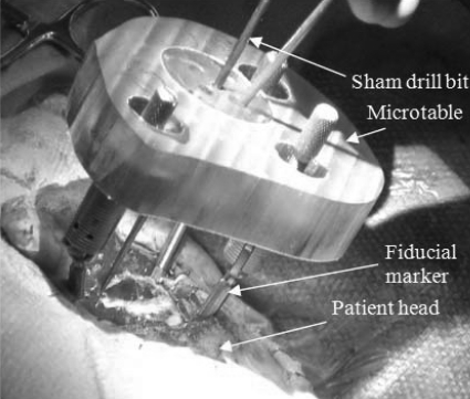
- Labadie R. F., Chodhury P., Cetinkaya E., Balachandran R., Haynes D. S., Fenlon M. R., Jusczyzck A. S., and Fitzpatrick J. F., “Minimally invasive, image-guided, facial-recess approach to the middle ear: Demonstration of the concept of percutaneous cochlear access in vitro,” Otol. Neurotol. 26(4), 557–562 (2005). 10.1097/01.mao.0000178117.61537.5b - DOI - PubMed
-
- Noble J. H., Majdani O., Labadie R. F., Dawant B., and Fitzpatrick J. M., “Automatic determination of optimal linear drilling trajectories for cochlear access accounting for drill-positioning error,” Int. J. Med. Robot. Comput. Assist. Surg. 6(3), 281–290 (2010). 10.1002/rcs.330 - DOI - PMC - PubMed
-
- Labadie R. F., Mitchell J., Balachandran R., and Fitzpatrick J. M., “Customized, rapid-production microstereotactic table for surgical targeting: Description of concept and in vitro validation,” Int. J. Comput. Assist. Radiol. Surg. 4(3), 273–280 (2009). 10.1007/s11548-009-0292-3 - DOI - PMC - PubMed
