. 2012 Feb;32(1):189-200.
doi: 10.1007/s10875-011-9603-x. Epub 2011 Oct 13.

Hemodialysis affects phenotype and proliferation of CD4-positive T lymphocytes

Affiliations

Hemodialysis affects phenotype and proliferation of CD4-positive T lymphocytes

Katarzyna A Lisowska et al. J Clin Immunol. 2012 Feb.

Abstract

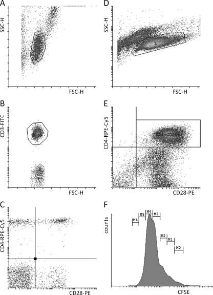
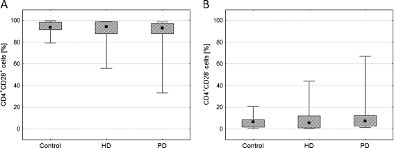
CD4(+) T lymphocytes of patients with chronic kidney disease (CKD) are characterized by reduced levels of crucial surface antigens and changes in the cell cycle parameters. Recombinant human erythropoietin (rhEPO) normalizes their altered phenotype and proliferative capacity. Mechanisms leading to the deficient responses of T lymphocytes are still not clear but it is postulated that immunological changes are deepened by hemodialysis (HD). Study of activation parameters of CD4(+) T lymphocytes in hemodialyzed and predialysis CKD patients could bring insight into this problem. Two groups of patients, treated conservatively (predialysis, PD) and hemodialyzed (HD), as well as healthy controls, were included into the study; neither had received rhEPO. Proportions of main CD4(+)CD28(+), CD4(+)CD25(+), CD4(+)CD69(+), CD4(+)CD95(+), and CD4(+)HLA-DR(+) lymphocyte subpopulations and proliferation kinetic parameters were measured with flow cytometry, both ex vivo and in vitro. No differences were seen in the proportions of main CD4(+) lymphocyte subpopulations (CD4(+)CD28(+), CD4(+)CD25(+), CD4(+)HLA-DR(+), CD4(+)CD69(+), CD4(+)CD95(+)) between all examined groups ex vivo. CD4(+) T lymphocytes of HD patients exhibited significantly decreased expression of co-stimulatory molecule CD28 and activation markers CD25 and CD69 after stimulation in vitro when compared with PD patients and healthy controls. HD patients showed also decreased percentage of CD4(+)CD28(+) lymphocytes proliferating in vitro; these cells presented decreased numbers of finished divisions after 72 h of stimulation in vitro and had longer G0→G1 time when compared to healthy controls. CD4(+) T lymphocytes of PD patients and healthy controls were characterized by similar cell cycle parameters. Our study shows that repeated hemodialysis procedure influences phenotype and proliferation parameters of CD4(+) T lymphocytes.


Figures

Fig. 1
Fig. 1
Flow cytometric analysis of subpopulations and proliferation of T lymphocytes ex vivo and in vitro. Whole blood cells were stained with monoclonal antibodies against CD3, CD4, and CD28 antigens. First, T lymphocytes were selected on the basis of their forward and side scatter characteristics (a) and CD3 expression (b). Finally, CD3+ T lymphocytes were visualized in dot-plot showing CD4 and CD28 (or other antigen) on the x- and y-axes, respectively (c). Meanwhile, proliferating T lymphocytes (stained with CFSE as described in “Patients and Methods”) were also selected on the basis of their forward and side scatter characteristics (d). Next, the subpopulation of lymphocytes of interest (for example CD4+CD28+ cells) was gated (e) and their divisions were shown in histogram (f)
Fig. 2
Fig. 2
Comparison of populations of main T lymphocytes ex vivo. Graphs show percentages of main populations: CD3+ (a), CD4+ (b), and CD8+ (c) as well as CD4/CD8 ratio (d) in all examined groups—healthy controls (Control), hemodialyzed patients (HD), and predialysis patients (PD). Midpoints of figures present medians, boxes present 25–75%, and whiskers outside present the minimum and maximum of all the data, Kruskal–Wallis test
Fig. 3
Fig. 3
Comparison of CD4+CD28+ and CD4+CD28 of subpopulations of T lymphocytes ex vivo. Graphs demonstrate the percentages of CD4+CD28+ (a) and CD4+CD28 (b) cells in all examined groups. Midpoints of figures present medians, boxes present 25–75%, and whiskers outside present the minimum and maximum of all the data, Kruskal–Wallis test
Fig. 4
Fig. 4
Comparison of subpopulations of main active CD4+ T lymphocytes ex vivo. Graphs demonstrate the percentages of CD4+CD69+ (a), CD4+CD25+ (b), CD4+CD95+ (c), and CD4+HLA-DR+ (d) cells in all examined groups. Midpoints of figures present medians, boxes present 25–75%, and whiskers outside present the minimum and maximum of all the data, Kruskal–Wallis test
Fig. 5
Fig. 5
Proportions of CD4+CD28+ and CD4+CD25+ T lymphocytes in vitro. Graphs present the percentages of CD4+CD28+ and CD4+CD25+ cells after 72 h (a, c) and 120 h (b, d) of stimulation with anti-CD3 antibody in examined groups. Midpoints of figures present medians, boxes present 25–75%, and whiskers outside present the minimum and maximum of all the data, *p < 0.05, Kruskal–Wallis test
Fig. 6
Fig. 6
Changes in the expression of CD28, CD69, and CD25 antigens on CD4+ T lymphocytes in vitro. Graphs show the expression of CD28, CD69, and CD25 antigens on CD4+ T lymphocytes after 72 h (a, c, e) and 120 h (b, d, f) of stimulation with anti-CD3 antibody in all examined groups. Midpoints of figures present medians, boxes present 25–75%, and whiskers outside present the minimum and maximum of all the data, *p < 0.05, Kruskal–Wallis test. MFI mean fluorescence intensity
Fig. 7
Fig. 7
Changes in main cell cycle parameters of CD4+CD28+ T lymphocytes. Graphs present the percentage of proliferating CD4+CD28+ cells after 72 h (a) and 120 h (b) of stimulation with anti-CD3 antibody, cell cycle duration (c), and time between phase G0 and G1 (d) in examined groups. Midpoints of figures present medians, boxes present 25–75%, and whiskers outside present the minimum and maximum of all the data, *p < 0.05, Kruskal–Wallis test
Fig. 8
Fig. 8
Comparison of the number of divisions of CD4+CD28+ T lymphocytes and their correlations with other cell cycle parameters. Graphs present the general number of divisions (a), the number of divisions per one cell (b), and the number of finished divisions (c) after 72 h of stimulation with anti-CD3 antibody in examined groups. Midpoints of ac present medians, boxes present 25–75%, and whiskers outside present the minimum and maximum of all the data, *p < 0.05, Kruskal–Wallis test. df Spearman R correlation
Fig. 9
Fig. 9
Comparison of main cell cycle parameters of CD4+CD28 T lymphocytes. Graphs present the percentage of proliferating CD4+CD28 cells (a), the general number of divisions (b), and the number of divisions per one cell (c) after 72 h of stimulation with anti-CD3 antibody in examined groups. Midpoints of ac present medians, boxes present 25–75%, and whiskers outside present the minimum and maximum of all the data, Kruskal–Wallis test

References

    1. Johnson CA, Levey AS, Coresh J, Levin A, Lau J, Eknoyan G. Clinical practice guidelines for chronic kidney disease in adults: part I. Definition, disease stages, evaluation, treatment, and risk factors. Am Fam Physician. 2004;70:869–876. - PubMed
    1. Herbelin A, Urena P, Zingraff J, Descamps-Latscha B. Influence of first long-term dialysis on uremia-associated increased basal production of interleukin-1 and tumor necrosis factor α by circulating monocytes. Nephrol Dial Transplant. 1991;6:349–357. - PubMed
    1. Cavaillon JM, Poignet JL, Fitting C, Delons S. Serum of interleukin-6 in long-term hemodialysis patients. Nephron. 1991;60:307–313. doi: 10.1159/000186770. - DOI - PubMed
    1. Ueki Y, Nagata M, Miyake S, Tominaga Y. Lymphocytes subsets in hemodialysis patients treated with recombinant human erythropoietin. J Clin Immunol. 1993;13:279–287. doi: 10.1007/BF00919387. - DOI - PubMed
    1. Morra L, Ponassi GA, Gurreri G, Moccia F, Mela GS, Bessone G. T lymphocyte subsets in chronic uremic patients treated with maintenance hemodialysis. Biomed Pharmacother. 1990;44:53–56. doi: 10.1016/0753-3322(90)90070-P. - DOI - PubMed

Publication types