. 2011 Oct 12;478(7368):225-8.
doi: 10.1038/nature10500.

Self-replication of information-bearing nanoscale patterns

Affiliations

Self-replication of information-bearing nanoscale patterns

Tong Wang et al. Nature. .

Abstract

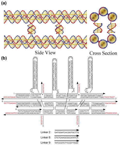
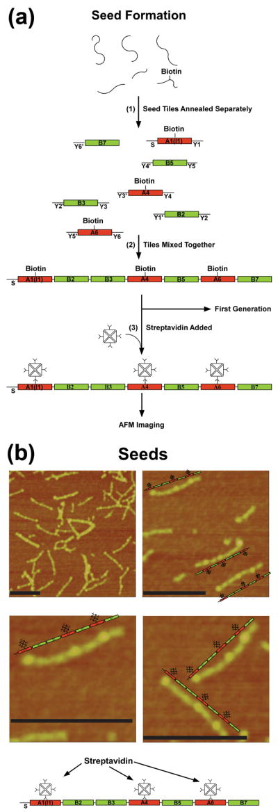
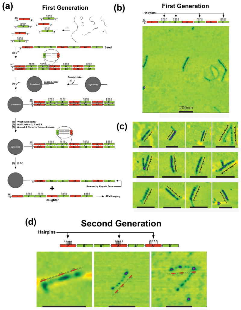
DNA molecules provide what is probably the most iconic example of self-replication--the ability of a system to replicate, or make copies of, itself. In living cells the process is mediated by enzymes and occurs autonomously, with the number of replicas increasing exponentially over time without the need for external manipulation. Self-replication has also been implemented with synthetic systems, including RNA enzymes designed to undergo self-sustained exponential amplification. An exciting next step would be to use self-replication in materials fabrication, which requires robust and general systems capable of copying and amplifying functional materials or structures. Here we report a first development in this direction, using DNA tile motifs that can recognize and bind complementary tiles in a pre-programmed fashion. We first design tile motifs so they form a seven-tile seed sequence; then use the seeds to instruct the formation of a first generation of complementary seven-tile daughter sequences; and finally use the daughters to instruct the formation of seven-tile granddaughter sequences that are identical to the initial seed sequences. Considering that DNA is a functional material that can organize itself and other molecules into useful structures, our findings raise the tantalizing prospect that we may one day be able to realize self-replicating materials with various patterns or useful functions.


Figures

Figure 1
Figure 1. DNA tile sequences and structures
(a) The P6HB Motif. This representation shows the way in which two BTX domains are paired by four lateral connections to form the P6HB motif. The cross section view shows two of the four helices that are formed by the lateral cohesive interactions. The interactions at the rear are eclipsed in this projection. (b) The Sequence and Structure of the B′ BTX Tile. Four helical domains, hairpins, are shown attached perpendicular to the BTX motif, so that they will create a topographic feature that can be detected in the atomic force microscope (AFM). Other tiles are shown in the Supplementary Information (S1).
Figure 2
Figure 2. DNA seeds
(a) Seed Formation. This drawing shows in step 1 how the individual strands of the seed tiles are self-assembled in separate vessels to produce seven different BTX tiles flanked by three sets of unique sticky ends labeled Y and a number; primed numbers are complementary to unprimed numbers. The red tiles are the A tiles and the green tiles are the B tiles. The A tiles contain a biotin group to enable their decoration by streptavidin. The tile labeled A1(I1) is the initiator tile. The strand labeled S on its left can bind to a dynabead during the replication process. Step 2 shows that these tiles produce 7-unit seeds when they are mixed together. The tiles are prepared for AFM imaging by the addition of streptavidin (Step 3) (b) AFM Images of Seeds. The image at the upper left shows a typical field slightly less than a square micron. Black scale bars correspond to 200nm. A large number of seeds are present, along with some multimeric complexes. The other three panels are zoomed images. A schematic image of each seed is shown next to the seed.
Figure 3
Figure 3. DNA generations
(a) Replication of the Seed Pattern in the First Generation. Strands are annealed in step 1, where the tiles are all flanked by the same connectors, designated Y and Z. The initiator tile contains a protected S-strand, paired with a cover strand, 6C. The B′ tiles contain the 4-hairpin markers for AFM imaging. In the presence of the seed tile (step 2), the strands assemble into a pattern mimicking the seed pattern. The magnetic dynabead is prepared in step 3, and attached to the seed (step 4). This is followed by a wash step, the addition of linkers and their annealing (steps 5-7). Heating the system to 37 °C results in the separation of the daughter 7-tile complex and the seed (removed magnetically). (b) Atomic Force Microscopy of First Generation Constructs Showing a Typical Field Slightly Larger than a Square Micron. Black scale bars correspond to 200nm. (c) Zoomed Images of Heptameric Daughter Complexes. These images, flanked by explanatory schematic images demonstrate that the ABBABAB pattern has been replicated successfully. (d) AFM Images of Second Generation Molecules. These zoomed images show the pattern that was programmed in the original seed tile.

References

    1. Lincoln TA, Joyce GF. Self-sustained replication of an RNA enzyme. Science. 2009;323:1229–1232. - PMC - PubMed
    1. Wintner EA, Conn MM, Rebek J., Jr Studies in molecular replication. Accts Chem Res. 1994;27:198–203.
    1. Schulman R, Winfree E. Synthesis of crystals with a programmable kinetic barrier to nucleation. Proc Nat Acad Sci (USA) 2007;104:15236–15241. - PMC - PubMed
    1. Lin C, et al. In vivo cloning of artificial DNA nanostructures. Proc Nat Acad Sci (USA) 2008;105:17626–17631. - PMC - PubMed
    1. Lee DH, Severin K, Yokobayashi Y, Ghadiri MR. Emergence of symbiosis in peptide self-replication through a peptide hypercyclic network. Nature. 1997;390:591–594. - PubMed

Publication types