

doi: 10.1002/mabi.201100180.
Epub 2011 Oct 12.
Pullulan hydrogels improve mesenchymal stem cell delivery into high-oxidative-stress wounds
Affiliations
- PMID: 21994074
- PMCID: PMC4157905
- DOI: 10.1002/mabi.201100180
Item in Clipboard
Pullulan hydrogels improve mesenchymal stem cell delivery into high-oxidative-stress wounds
Macromol Biosci.
.
Abstract
Cell-based therapies for wound repair are limited by inefficient delivery systems that fail to protect cells from the acute inflammatory environment. Here, a biomimetic hydrogel system is described that is based on the polymer pullulan, a carbohydrate glucan known to exhibit potent antioxidant capabilities. It is shown that pullulan hydrogels are an effective cell delivery system and improve mesenchymal stem cell survival and engraftment in high-oxidative-stress environments. The results suggest that glucan hydrogel systems may prove beneficial for progenitor-cell-based approaches to skin regeneration.
Copyright © 2011 WILEY-VCH Verlag GmbH & Co. KGaA, Weinheim.
Figures

Hydrogel delivery of MSCs. (A) Molecular structure of pullulan. (B) Antioxidant capacity of the pullulan polymer as measured using an ABTS assay. n = 3 for each data point. Statistical significance is relative to the spectrophotometric absorbance of hydrogen peroxide-free solution at the respective concentrations. (C) Antioxidant properties of the pullulan hydrogel system as measured using the Amplex Red assay kit. Incubation with the pullulan hydrogel decreased the amount of hydrogen peroxide at 30, 60, 120, and 240 min. Significance values are relative to control at the same concentration. (D) 1H NMR demonstrates increasing signal at δ = 8.46 in pullulan hydrogels following exposure to hydrogen peroxide, consistent with the oxidation of primary alcohol groups at C6. (E) Graph showing the increase in oxidation (aldehyde peak) of pullulan hydrogels over time, as measured by calculating the area under the signal at δ = 8.46. In contrast, dextran hydrogels (which lack a free C6 primary hydroxyl) exhibit less antioxidant capacity in this assay. Data are representative of three independent experiments. (F) Scanning electron micrographs demonstrating the differential morphology of plated MSCs (top row) compared to hydrogel-seeded MSCs (bottom row). Clusters of MSCs are shown within the three-dimensional environment of the porous hydrogel matrix. Scale bar 100 μm (left) and 15 μm (right). Values represent means ± SD. *p <0.05.
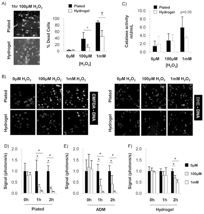
Pullulan-based hydrogels protect MSCs from ROS-induced damage. (A) Representative images (left) and quantification (right) of live/dead assay 1 h following exposure to hydrogen peroxide. n = 6. (B) Representative images of intracellular ROS activity as detected with CMFDA and DHE. Oxidized products are unable to be secreted from MSCs and are detected by wavelength-specific fluorescence. (C) Catalase activity of MSCs following 1 h exposure to hydrogen peroxide. n = 3. (D–F) Quantification of in vitro bioluminescence imaging of luc + MSCs seeded onto (D) standard tissue culture plates, (E) decellularized human dermis, and (F) pullulan-based hydrogels. n = 4. Values represent means ± SD. *p <0.05, †p <0.01.

Subcutaneous delivery of MSCs using pullulan hydrogels. (A) Representative images and (B) quantification of bioluminescent imaging of luc + MSCs delivered subcutaneously via injection (top row) or hydrogel scaffold (bottom row). Data are relative to bioluminescence at day 1 for the respective conditions. (C) Representative image of luc/eGFP + MSCs at day 10 post-injury delivered via hydrogel. Delivered MSCs were distributed homogeneously throughout the provisional dermal matrix as opposed to a subcutaneous location. Scale bar 20 μm. n = 4. Values represent means ± SD. *p <0.05.
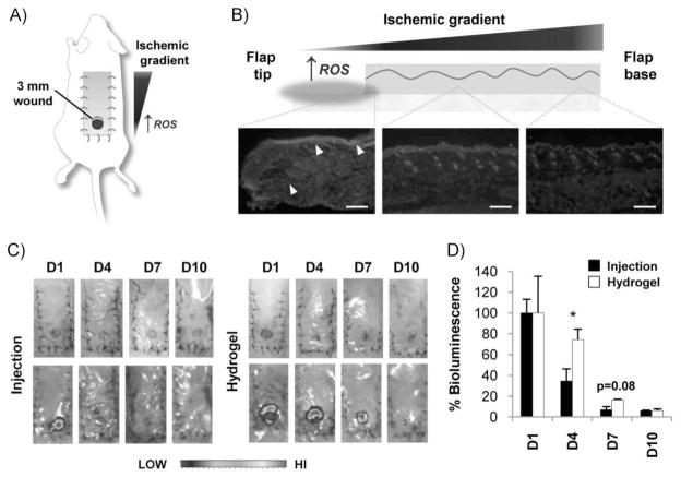
Delivery of MSCs into ischemic excisional wounds. (A) Schematic of ischemic excisional wound model. An ischemic gradient is established with lowest levels of oxygen tension at the distal tip of the peninsular flap. (B) DHE immunofluoresence demonstrating highest levels of ROS at the ischemic flap tip. Arrowheads point to elevated ROS in the epithelial layer and dermal wound margin. Images were taken from the same flap at the same exposure from the distal, middle, and proximal third that corresponds to the left, middle, and right micrographs. Scale bar 200 μm. (C) Gross photographs (top) and IVIS bioluminescence imaging (bottom) of the ischemic excisional wound model following injection or hydrogel delivery of MSCs. (D) Quantification of bioluminescent imaging of luc + MSCs delivered via local injection or hydrogel scaffold. Data are relative to bioluminescence at day 1 for the respective conditions. n = 4. Values represent means ± SEM. *p <0.05.
References
-
- Gurtner GC, Werner S, Barrandon Y, Longaker MT. Nature. 2008;453:314. - PubMed
-
- Singer AJ, Clark RA. N Engl J Med. 1999;341:738. - PubMed
-
- Schafer M, Werner S. Pharmacol Res. 2008;58:165. - PubMed
-
- Khodr B, Khalil Z. Free Radic Biol Med. 2001;30:1. - PubMed
-
- Callaghan MJ, Ceradini DJ, Gurtner GC. Antioxid Redox Signal. 2005;7:1476. - PubMed
Publication types
MeSH terms
Substances
Grants and funding
LinkOut - more resources
Full Text Sources
Other Literature Sources
Medical
