. 2012 Feb;93(Pt 2):235-246.
doi: 10.1099/vir.0.034033-0. Epub 2011 Oct 12.

Hepatitis C virus activates interleukin-1β via caspase-1-inflammasome complex

Affiliations

Hepatitis C virus activates interleukin-1β via caspase-1-inflammasome complex

Dylan Burdette et al. J Gen Virol. 2012 Feb.

Retraction in

Expression of concern in

Abstract

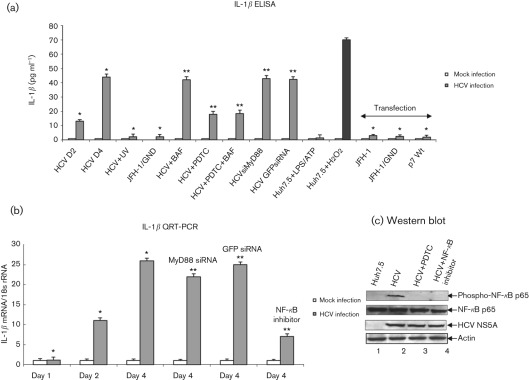
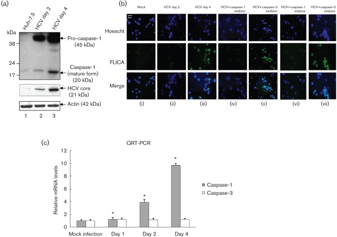
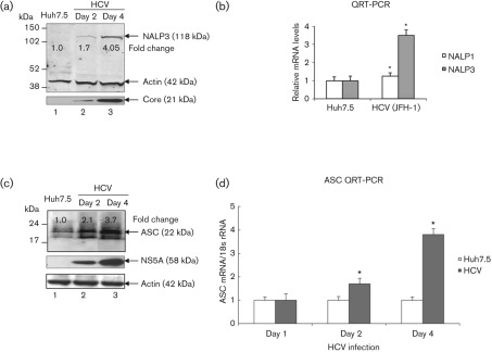
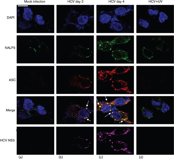
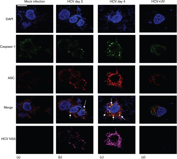
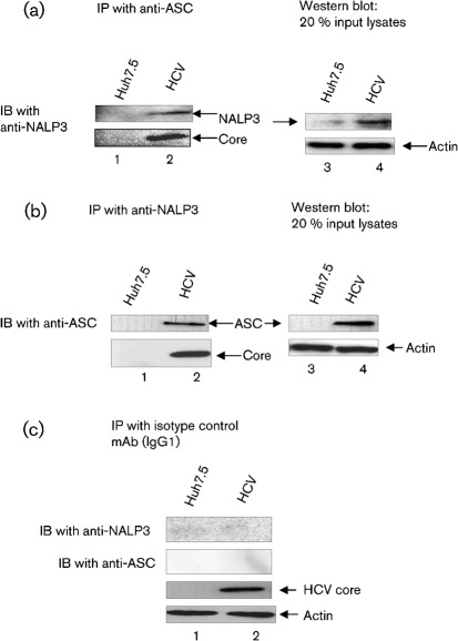
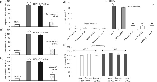
Interleukin-1β (IL-1β) is a potent pro-inflammatory cytokine involved in the pathogenesis of HCV, but the sensors and underlying mechanisms that facilitate HCV-induced IL-1β proteolytic activation and secretion remains unclear. In this study, we have identified a signalling pathway leading to IL-1β activation and secretion in response to HCV infection. Previous studies have shown the induction and secretion of IL-1β through the inflammasome complex in macrophages/monocytes. Here, we report for the first time the induction and assembly of the NALP3-inflammasome complex in human hepatoma cells infected with HCV (JFH-1). We demonstrate that activation of IL-1β in HCV-infected cells involves the proteolytic processing of pro-caspase-1 into mature caspase-1 in a multiprotein inflammasome complex. Next, we demonstrate that HCV is sensed by NALP3 protein, which recruits the adaptor protein ASC for the assembly of the inflammasome complex. Using a small interfering RNA approach, we further show that components of the inflammasome complex are involved in the activation of IL-1β in HCV-infected cells. Our study also demonstrates the role of reactive oxygen species in HCV-induced IL-1β secretion. Collectively, these observations provide an insight into the mechanism of IL-1β processing and secretion, which is likely to provide novel strategies for targeting the viral or cellular determinants to arrest the progression of liver disease associated with chronic HCV infection.


Figures

Fig. 1.
Fig. 1.
Activation of IL-1β by HCV. (a) Huh7.5 cells were incubated with HCV cell culture supernatant (m.o.i. of 1) for 6 h, and cultured for 2 and 4 days. Huh7.5 cells were incubated with UV-irradiated HCV, or supernatant from cells expressing JFH-1/GND for 6 h and cultured for 4 days. Huh7.5 cells were incubated with LPS (20 ng ml−1 for 6 h) followed by ATP (5 mM for 20 min) and H2O2 (500 µM for 20 min). HCV-infected cells at day 4 were incubated with BAF (20 nM for 12 h) and/or PDTC (100 µM for 6 h). HCV-infected cells were transfected with MyD88 siRNA or GFP siRNA for 4 days. Huh7.5 cells were also transfected with in vitro-transcribed JFH-1, JFH-1/GND RNA for 10 h, or plasmid expressing HCV p7 protein for 4 days. Cells were serum starved for 4 h, and cell culture supernatants were collected, centrifuged and subjected to IL-1β ELISA. The data shown here represent the means+sd of at least three independent experiments performed in triplicate. *, P<0.05 compared with mock-infected cells. **, P<0.05 compared with HCV-infected cells at day 4. (b) Induction of IL-1β mRNA by HCV. Huh7.5 cells were incubated with HCV cell culture supernatant as described above. HCV-infected cells were incubated with NF-κB inhibitor (20 µM for 6 h), PDTC (100 µM for 6 h) and transfected with MyD88 or GFP siRNA as described in Methods. Cells were serum starved for 4 h and total cellular RNA was extracted and subjected to cDNA synthesis. QRT-PCR was carried out using SYBR green master mix (ABI) and IL-1β-specific primer sets. 18s rRNA was used as an internal control. The values represent the means+sd of at least three independent experiments performed in triplicate. *, P<0.05 compared with mock-infected control cells. **, P<0.05 compared with HCV-infected cells at day 4. (c) Activation of NF-κB by HCV. Equal amounts of cellular lysates from Huh7.5, HCV-infected cells, and treated with PDTC or NF-κB inhibitor were Western blotted with indicated antibodies. Lanes 1 and 2, lysates from Huh7.5 cells and HCV-infected cells; lanes 3 and 4, HCV-infected cells treated with PDTC or NF-κB inhibitor, respectively. Bottom panels represent the expression of HCV NS5A and actin controls.
Fig. 2.
Fig. 2.
Activation of caspase-1 by HCV. (a) Mock-infected and HCV-infected Huh7.5 cells were serum starved for 4 h and cellular lysates were subjected to SDS-PAGE followed by Western blot analysis using caspase-1 antibody. Lane 1, Huh7.5 lysates; lanes 2 and 3, lysates from Huh7.5 cells infected with HCV. Bottom panels represent the expression of HCV core and actin controls. (b) FLICA assay. Mock-infected and HCV-infected cells were serum starved for 4 h and treated with 50 µM caspase-1 inhibitor (z-VAD-fmk) for 1 h (iv), and 2 h (vi) or 100 µM caspase-3 inhibitor (DEVD) for 1 h (v), and 2 h (vii). The cells were incubated with the FLICA reagent and Hoescht nuclear stain for 1 h, before being visualized via fluorescence microscopy. (c) Total cellular RNA from mock-infected and HCV-infected cells were subjected to QRT-PCR using caspase-1-specific primers. The data shown here represent the means+sd of at least three independent experiments performed in triplicate. *, P<0.05 compared with mock-infected control cells.
Fig. 3.
Fig. 3.
Induction of NALP3 and ASC expression by HCV. (a) Total cellular lysates from mock-infected and HCV-infected cells were subjected to Western blot analysis using antibodies against NALP3, HCV core and actin. Lane 1, Huh7.5 lysates; lanes 2 and 3, lysates from HCV-infected cells. (b) Total cellular RNA was extracted from mock-infected and HCV-infected cells at day 4 and subjected to QRT-PCR using NALP1- and NALP3-specific primers. 18S rRNA was used as an internal control. The data shown here represent the means+sd of at least three independent experiments performed in triplicate. *, P<0.05 compared with mock-infected control cells. (c) Induction of ASC by HCV. Total cellular lysates were Western blotted using antibodies against ASC, HCV NS5A and actin. Lane 1, Huh7.5 lysates; lanes 2 and 3, lysates from HCV-infected cells. (d) Total cellular RNA from mock-infected and HCV-infected cells were subjected to QRT-PCR using ASC-specific primers. The values represent the means+sd of at least three independent experiments performed in triplicate. *, P<0.05 compared with mock-infected cells.
Fig. 4.
Fig. 4.
HCV infection activates the inflammasome complex. Mock-infected and HCV-infected cells at indicated time points were serum starved for 4 h and incubated with NALP3, ASC and HCV NS3 antibodies for 1 h at RT, followed by incubation with secondary antibodies for NALP3 (anti-rabbit Alexa Fluor 488), ASC (anti-goat Alexa Fluor 546), NS3 (anti-mouse Alexa Fluor 633). DAPI was used as a nuclear stain. Arrows represent the staining and colocalization of ASC with NALP3. Bar, 10 µm.
Fig. 5.
Fig. 5.
Similar to Fig. 4, mock-infected and HCV-infected cells were stained with ASC, caspase-1 and HCV NS3 antibodies for 1 h at RT, followed by incubation with secondary antibodies for ASC (anti-goat Alexa Fluor 546), caspase-1 (anti-rabbit Alexa Fluor 488) and NS3 (anti-mouse Alexa Fluor 633). Arrows represent the staining and colocalization of ASC with caspase-1. Bar, 10 µm.
Fig. 6.
Fig. 6.
Interaction of ASC with NALP3. Mock-infected and HCV-infected cells were cultured for 4 days and serum starved for 4 h. Equal amounts (500 µg) of cellular lysates from mock-infected and HCV-infected cells were immunoprecipitated with anti-ASC (a), anti-NALP3 (b) or isotype control antibodies (c), and immunoblotted with anti-NALP3 or anti-ASC antibodies. Lane 1, mock-infected lysates; lane 2, HCV-infected lysates. Bottom panels represent the expression of HCV core protein and actin protein loading controls. Lane 4 in (a) and (b) represent 20 % input lysates from mock-infected and HCV-infected cells.
Fig. 7.
Fig. 7.
Effect of NALP3, ASC and caspase-1 siRNA on IL-1β secretion. (a–c) Mock-infected and HCV-infected cells were transfected with siRNA against GFP, NALP3, ASC and caspase-1 as described in Methods. At day 4, total cellular RNA was extracted using TRIzol, cDNA was synthesized and QRT-PCR was performed using GFP-, NALP3-, ASC- and caspase-1-specific primers (Supplementary Table S1). 18S rRNA was used as an internal control. The data shown here represent the means+sd of at least three independent experiments performed in triplicate. *, P<0.05 compared with GFP siRNA-transfected control cells. (d) Effect of NALP3, ASC and caspase-1 siRNA on IL-1β secretion. Cell culture supernatants from siRNA-transfected mock-infected and HCV-infected cells were subjected to IL-1β ELISA. *, P<0.05 compared with mock-infected control cells. **, P<0.05 compared with GFP siRNA-transfected cells. (e) Cytotoxicity assay. Equal volume of cell culture medium from mock-infected and HCV-infected cells, transfected with GFP, caspase-1 and NALP3 siRNA were subjected to cytotoxicity assay using CytoTox-ONE. RFU, Relative fluorescence unit.

References

    1. Allen I. C., Scull M. A., Moore C. B., Holl E. K., McElvania-TeKippe E., Taxman D. J., Guthrie E. H., Pickles R. J., Ting J. P. (2009). The NLRP3 inflammasome mediates in vivo innate immunity to influenza A virus through recognition of viral RNA. Immunity 30, 556–56510.1016/j.immuni.2009.02.005 - DOI - PMC - PubMed
    1. Bartenschlager R., Lohmann V. (2000). Replication of hepatitis C virus. J Gen Virol 81, 1631–1648 - PubMed
    1. Blight K. J., Kolykhalov A. A., Rice C. M. (2000). Efficient initiation of HCV RNA replication in cell culture. Science 290, 1972–197410.1126/science.290.5498.1972 - DOI - PubMed
    1. Blight K. J., McKeating J. A., Rice C. M. (2002). Highly permissive cell lines for subgenomic and genomic hepatitis C virus RNA replication. J Virol 76, 13001–1301410.1128/JVI.76.24.13001-13014.2002 - DOI - PMC - PubMed
    1. Bryan N. B., Dorfleutner A., Rojanasakul Y., Stehlik C. (2009). Activation of inflammasomes requires intracellular redistribution of the apoptotic speck-like protein containing a caspase recruitment domain. J Immunol 182, 3173–318210.4049/jimmunol.0802367 - DOI - PMC - PubMed

Publication types

LinkOut - more resources