. 2011 Dec;85(24):13105-13.
doi: 10.1128/JVI.05598-11. Epub 2011 Oct 12.

A virus-like particle-based Epstein-Barr virus vaccine

Affiliations

A virus-like particle-based Epstein-Barr virus vaccine

Romana Ruiss et al. J Virol. 2011 Dec.

Abstract

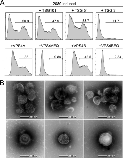
Epstein-Barr Virus (EBV) is an ubiquitous human herpesvirus which can lead to infectious mononucleosis and different cancers. In immunocompromised individuals, this virus is a major cause for morbidity and mortality. Transplant patients who did not encounter EBV prior to immunosuppression frequently develop EBV-associated malignancies, but a prophylactic EBV vaccination might reduce this risk considerably. Virus-like particles (VLPs) mimic the structure of the parental virus but lack the viral genome. Therefore, VLPs are considered safe and efficient vaccine candidates. We engineered a dedicated producer cell line for EBV-derived VLPs. This cell line contains a genetically modified EBV genome which is devoid of all potential viral oncogenes but provides viral proteins essential for the assembly and release of VLPs via the endosomal sorting complex required for transport (ESCRT). Human B cells readily take up EBV-based VLPs and present viral epitopes in association with HLA molecules to T cells. Consequently, EBV-based VLPs are highly immunogenic and elicit humoral and strong CD8+ and CD4+ T cell responses in vitro and in a preclinical murine model in vivo. Our findings suggest that VLP formulations might be attractive candidates to develop a safe and effective polyvalent vaccine against EBV.


Figures

Fig. 1.
Fig. 1.
293-VII+ cells release VLPs with a B cell tropism that lack viral DNA. (A) B cells were isolated from human adenoids and incubated for 2 days with VLPs from 293-VII+ cells (bold line) or EBV 2089 (fine line). VLPs from 293-VII+ cells render cells faintly GFP positive, indicating a direct interaction. Incubation of B cells with EBV 2089 stocks results in a majority of cells which are dimly GFP positive and 12.5% strongly GFP-positive cells in which the gfp gene is expressed after viral transduction. (B) VLPs from 293-VII+ cells contain the exosome markers hsp70 and tsg101 and the EBV structural proteins BLLF1 (gp350/220), BNRF1 (gp140), and BALF4 (gp125). Lysates from VLPs from 293-VII+ cells and exosomes from HEK293 were spotted onto nitrocellulose and hybridized to the antibodies indicated. (C) VLPs released from induced 293-VII+ cells exclusively bind to human CD21-positive cells. PBMCs were treated either with GFP-negative exosomes from parental HEK293 cells (left dot blot), with exosomes spontaneously released from uninduced 293-VII+ cells (middle dot blot), or with VLPs from lytically induced 293-VII+ cells (right dot blot) and analyzed by flow cytometry. Exosomes from uninduced 293-VII+ cells bound weakly and unspecifically to PBMCs (inset). In contrast, VLPs have a CD21 tropism, as revealed by GFP-positive CD21+ cells (right dot blot). (D) VLPs interact with B cells via gp350. Human primary B cells were incubated overnight with VLPs either alone or in the presence of inhibitory gp350- or CD21-specific antibodies. Binding of VLPs to B cells was quantified by flow cytometry by measuring GFP. Both antibodies interfered with VLP binding. (E) VLPs from 293-VII+ cells were incubated with primary B cells, and the interaction was analyzed by laser scanning microscopy, revealing a colocalization of CD21 (green) and gp350 (red) in three-dimensional reconstruction images. Cells were counterstained with 4′,6-diamidino-2-phenylindole (blue). (F) BZLF1- and BRLF1-specific PCR analysis detected viral mRNA in virions isolated from induced EBV 2089-infected cells and in VLPs from induced 293-VII+ cells. In contrast, viral DNA was detectable in EBV 2089-infected cells only. WT, wild type; RNA, PCR performed without reverse transcription proved that the RNA preparation was free of contaminating DNA.
Fig. 2.
Fig. 2.
EBV uses the exosome biogenesis pathway for viral egress. (A) HEK293 producer cells stably transfected with wild-type EBV 2089 (8) were transiently cotransfected with p509, an expression plasmid for BZLF1, to induce EBV's lytic cycle, together with expression plasmids encoding TSG101 or VSP4 proteins or dominant-negative mutants thereof. The supernatants were collected 3 days later and used for the infection of Raji cells. GFP-positive Raji cells were quantified by flow cytometry 3 days later. The numbers indicate the percentage of EBV-infected Raji cells. It became obvious that the dominant-negative mutants VPS4AEQ, VPS4BEQ, and TSG101 3′ completely blocked the release of infectious wild-type EBV 2089. (B) Electron microscopy revealed that purified VLPs from 293-VII+ cells are round and approximately 100 nm in diameter and morphologically resemble infectious EBV virions (27).
Fig. 3.
Fig. 3.
VLP-loaded B cells are efficient stimulators of autologous T cells. (A) Overnight incubation of CD19+ B cells purified from PBMCs with 293-VII+-derived VLPs induced the surface expression of the costimulatory molecules ICAM-1/CD54, B7.1/CD80, and B7.2/CD86 (black line) compared to B cells incubated with exosomes from HEK293 cells (gray region). (B) VLP-loaded B cells are potent stimulators of a BNRF1-specific CD4+ T cell clone. A mini-LCL line (34) was loaded with serial dilutions of VLPs obtained from lytically induced 293-VII+ cells. The loaded cells were used as stimulators for an autologous CD4+ T cell clone, which recognizes a BNRF1-specific epitope (32). Stimulation of the T cell clone with the mini-LCL line loaded with exosomes (exo) from HEK293 cells was used as a negative control, and an autologous LCL, which expresses BNRF1, was used as a positive control. (C) Reactivation of EBV-specific T lymphocytes with VLPs depends on CD19+ B cells. CD3+ T cells from a healthy donor who had been immunized with Twinrix 2 years previously were stimulated three times within a period of 14 days with autologous, lethally irradiated stimulator cells, as indicated, which had been loaded with HEK293 exosomes (exo), VLPs from 293-VII+ cells, or 30 μl Twinrix (corresponding to 22 ELISA units inactivated HAV and 0.6 μg HBV surface protein). The stimulator cells were either unfractionated PBMCs, marker-assisted congenic screening-sorted CD19+ B cells, or PBMCs from which the B cells were depleted (CD19). After three rounds of stimulation, reactivation of T cells was assessed in an IFN-γ ELISPOT assay, using autologous PBMCs loaded with either VLPs, exosomes, or Twinrix as targets. VLP-induced T cell reactivation was strictly dependent on the presence of CD19+ B cells as stimulators. Shown are a scan of the ELISPOT assay and the calculated numbers.
Fig. 4.
Fig. 4.
VLPs from 293-VII+ cells selectively expand EBV-specific CD4+ and CD8+ T cells. PBMCs from different EBV-positive donors were lethally irradiated, loaded with either VLPs from lytically induced 293-VII+ cells or exosomes from HEK293 cells, or left untreated and used as stimulators for autologous PBMCs. After 28 days and three rounds of stimulation, cells were analyzed by flow cytometry. (A) VLP- but not exosome-loaded (exo) or untreated (w/o) irradiated PBMCs expanded CD4+ T cells as described recently (2). (B) PBMCs loaded with VLPs reactivated and expanded EBV-specific CD8+ T cells, as revealed by staining with HLA B03/B08- or A02-restricted tetramers/pentamers to selected EBV protein epitopes. A tetramer (KIF) to the cellular protein Her2/neu served as a negative control. (C) VLP-loaded irradiated PBMCs reliably expanded EBV-specific CD8+ cells from four different donors up to 30-fold within 28 days, whereas the total number of CD8+ T cells dropped by about half compared to the initial CD8+ T cell numbers.
Fig. 5.
Fig. 5.
VLPs elicit EBV-specific humoral and cellular immune responses in immunized mice. BALB/c mice were immunized twice with 10 μg VLPs from 293-VII+ cells (n = 4) or with the same amounts of exosomes (exo) from HEK293 cells (n = 2). Sera and splenocytes were analyzed 4 weeks after the last immunization. (A) In ELISAs, sera from VLP-immunized mice (black bars) but not from exosome-immunized mice (gray bars) contain antibodies specific to EBV proteins present in virions (24). OD, optical density. (B) Antibodies generated in mice immunized with VLPs from 293-VII+ cells neutralize infectious EBV. GFP-positive EBV 2089 stocks were preincubated for 30 min with sera from mice immunized with VLPs or exosomes and subsequently used to infect human primary B cells at a calculated multiplicity of infection of 0.1. Forty-eight hours later, the number of GFP-expressing infected cells was determined by flow cytometry. The neutralizing anti-gp350 antibody 72A1 was used as a positive control at two different concentrations. (C) VLPs from 293-VII+ cells activate EBV-specific T cells. The occurrence of EBV-specific T cells in mice immunized with VLPs (black bars) or exosomes (gray bars) was measured in a mouse-specific IFN-γ ELISPOT assay with irradiated splenocytes as APCs loaded with lysates from HEK293 cells which had been transiently transfected with expression plasmids encoding the indicated viral proteins. The CMV protein pp65 was used to control the specificity of the assays.

Similar articles

Cited by

References

    1. Ablashi D. V., et al. 1997. Epstein-Barr virus and Kaposi's sarcoma herpesvirus/human herpesvirus 8, p. In (ed.), IARC monograph on the evaluation of carcinogenic risks to humans. International Association for Research on Cancer, Lyon, France
    1. Adhikary D., Behrends U., Feederle R., Delecluse H. J., Mautner J. 2008. Standardized and highly efficient expansion of Epstein-Barr virus-specific CD4+ T cells by using virus-like particles. J. Virol. 82:3903–3911 - PMC - PubMed
    1. Adhikary D., et al. 2006. Control of Epstein-Barr virus infection in vitro by T helper cells specific for virion glycoproteins. J. Exp. Med. 203:995–1006 - PMC - PubMed
    1. Ahlers J. D., Belyakov I. M. 2010. Memories that last forever: strategies for optimizing vaccine T-cell memory. Blood 115:1678–1689 - PMC - PubMed
    1. Altmann M., Hammerschmidt W. 2005. Epstein-Barr virus provides a new paradigm: a requirement for the immediate inhibition of apoptosis. PLoS Biol. 3:e404. - PMC - PubMed

Publication types

MeSH terms