. 2011 Dec;85(24):13398-408.
doi: 10.1128/JVI.06250-11. Epub 2011 Oct 12.

Structural characterization of the viral and cRNA panhandle motifs from the infectious salmon anemia virus

Affiliations

Structural characterization of the viral and cRNA panhandle motifs from the infectious salmon anemia virus

Robert G Brinson et al. J Virol. 2011 Dec.

Abstract

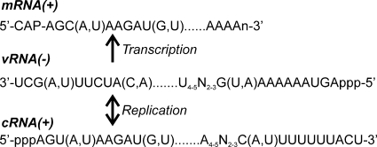
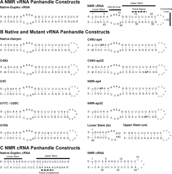
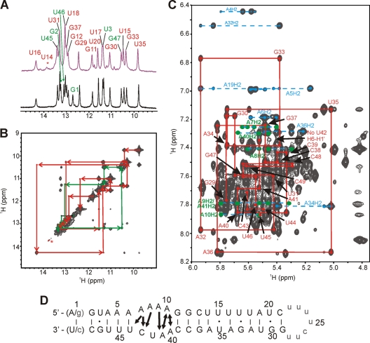
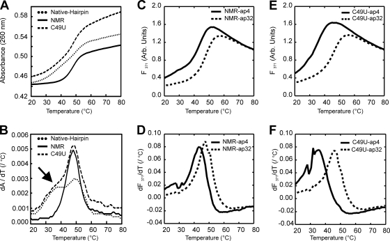
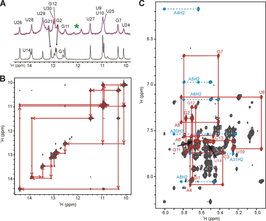
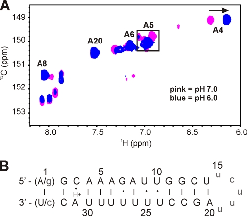
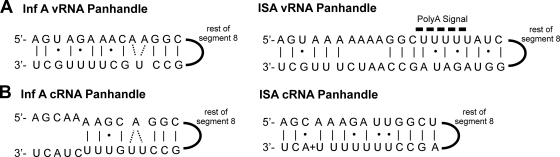
Infectious salmon anemia virus (ISAV) has emerged as a virus of great concern to the aquaculture industry since it can lead to highly contagious and lethal infections in farm-raised salmon populations. While little is known about the transcription/replication cycle of ISAV, initial evidence suggests that it follows molecular mechanisms similar to those found in other orthomyxoviruses, which include the highly pathogenic influenza A (inf A) virus. During the life cycle of orthomyxoviruses, a panhandle structure is formed by the pairing of the conserved 5' and 3' ends of each genomic RNA. This structural motif serves both as a promoter of the viral RNA (vRNA)-dependent RNA polymerase and as a regulatory element in the transcription/replication cycle. As a first step toward characterizing the structure of the ISAV panhandle, here we have determined the secondary structures of the vRNA and the cRNA panhandles on the basis of solution nuclear magnetic resonance (NMR) and thermal melting data. The vRNA panhandle is distinguished by three noncanonical U · G pairs and one U · U pair in two stem helices that are linked by a highly stacked internal loop. For the cRNA panhandle, a contiguous stem helix with a protonated C · A pair near the terminus and tandem downstream U · U pairs was found. The observed noncanonical base pairs and base stacking features of the ISAV RNA panhandle motif provide the first insight into structural features that may govern recognition by the viral RNA polymerase.


Figures

Fig. 1.
Fig. 1.
Schematic for synthesis of mRNA and replication of the viral RNA of all eight segments (consensus sequence shown) from ISAV (18, 28). The mRNA is defined by a 5′ catabolite gene activator protein (CAP) structure that is derived from cellular mRNA and a 3′ poly(A) tail. In contrast, the cRNA from which copies of the genomic vRNA are made does not have these modifications.
Fig. 2.
Fig. 2.
ISAV panhandle sequences from segment 8. (A and B) Native and mutant vRNA panhandle constructs used for NMR analysis (A) and thermal melting analysis (B). AP, positions where adenosine was substituted with 2-aminopurine. (C) cRNA panhandle constructs used for NMR analysis. Nonnative bases are in lowercase. Canonical base pairing is indicated by solid lines. In panel B, base changes made relative to the NMR vRNA construct are in bold.
Fig. 3.
Fig. 3.
NMR characterization of the ISAV vRNA panhandle from segment 8. (A) Imino proton region of 1D Watergate at 10°C. Upper spectrum, 23-mer native duplex vRNA with the native A-U terminal pair; lower spectrum, final NMR vRNA hairpin construct (49-mer) with a G-C terminal pair. Sequential imino proton assignments of the lower stem are given in green, and the upper stem assignments are in red and were determined by the 2D NOESY in panel B. (B) Imino proton region of a 2D NOESY-Watergate spectrum (τmix = 50 ms) at 10°C in 3% 2H2O for the NMR vRNA panhandle construct. The imino assignments are listed on the 1D projection. (C) Aromatic-anomeric region of a 2D NOESY spectrum (τmix = 250 ms) at 20°C in 99.96% 2H2O. A representative sequential NOE walk, H6/H8-H1′, from G29 to C49 is shown as a red trace. The NOE walk from G1 to C21 is not shown for clarity. Adenosine H2-H1′ NOE correlations are shown in cyan. Adenosine H2-H1′ NOEs in the internal loop are highlighted in green. (D) Summary of vRNA secondary structure determined from the NMR data. As discussed in the text, both native A-U and nonnative G-C terminal pairs were used in NMR constructs. Solid lines and dots, canonical and noncanonical base pairing, respectively. Nonnative bases are in lowercase. The arrows represent observed interstrand H2-H1′ NOEs within the internal loop.
Fig. 4.
Fig. 4.
UV and fluorescence melting data for vRNA panhandle constructs. (A and B) UV-detected melts (A) and first-derivative plots (B) of native hairpin, NMR vRNA, and C49U vRNA panhandles. Arrow, the shoulder observed for the melt of the lower stem in the C49U vRNA panhandle. (C and D) Fluorescence-detected melts (C) and first-derivative plots (D) for the NMR-ap4 and-ap32 vRNA panhandle constructs. (E and F) Fluorescence-detected melts (E) and first-derivative plots (F) for the C49U-ap4 and-ap32 vRNA panhandle constructs. Melting temperatures derived from the melting transitions are given in Table 1.
Fig. 5.
Fig. 5.
NMR characterization of ISAV cRNA panhandle. (A) Imino proton region of 1D Watergate at 10°C and pH 6.0. Upper spectrum, 23-mer native duplex cRNA with the native A-U terminal pair and the upper stem; lower spectrum, final NMR cRNA construct (33-mer) with a G-C terminal pair. Imino proton assignments were determined by the 2D NOESY in panel B. Green asterisk, minor peaks from insignificant base-pairing interactions in the upper stem of the native duplex cRNA. See the text for details. (B) Imino proton region of a 2D NOESY-Watergate spectrum (τmix = 150 ms) at 10°C in 3% 2H2O. Sequential imino proton assignments are given in red. (C) Aromatic-anomeric region of a 2D NOESY spectrum (τmix = 250 ms) at 20°C in 99.96% 2H2O. An NOE walk, H6/H8-H1′, from G1 to U14 is shown as a red trace. The NOE walk from U15 to C33 is not shown for clarity. Adenosine H2-H1′ correlations are shown in cyan.
Fig. 6.
Fig. 6.
(A) Overlay of adenosine H2-C2 region of a 1H-13C constant-time HSQC spectrum of a 13C/15N A/U-labeled NMR cRNA panhandle at 20°C. The proton carrier frequency was set to 4.71 ppm. Listed assignments were made at pH 6.0. The boxed region indicates the coalescing of 4 peaks into one peak from pH 7.0 to 6.0. An arrow indicates the upfield shift of A4 after the pH is dropped to pH 6.0. (B) Summary of cRNA secondary structure as determined from the NMR data. As discussed in the text, both native A-U and nonnative G-C terminal pairs were used in the NMR constructs. Solid lines and dots, canonical and noncanonical base pairings, respectively; H+, the protonated A31. Nonnative bases are in lowercase.
Fig. 7.
Fig. 7.
Schematic representations of the NMR-determined secondary structures for the segment 8 vRNA panhandle motifs from influenza A/PR/8/34 virus (2) and ISAV (A) and segment 8 cRNA panhandle motifs from influenza A/PR/8/34 virus (49) and ISAV (B). Solid lines and dots, canonical and noncanonical base pairings, respectively; solid semicircle, the rest of genomic segment 8.

Similar articles

Cited by

References

    1. Acharya S., et al. 2004. Significant pKa perturbation of nucleobases is an intrinsic property of the sequence context in DNA and RNA. J. Am. Chem. Soc. 126:8674–8681 - PubMed
    1. Bae S. H., et al. 2001. Structural features of an influenza virus promoter and their implications for viral RNA synthesis. Proc. Natl. Acad. Sci. U. S. A. 98:10602–10607 - PMC - PubMed
    1. Batey R. T., Battiste J. L., Williamson J. R. 1995. Preparation of isotopically enriched RNAs for heteronuclear NMR. Methods Enzymol. 261:300–322 - PubMed
    1. Bouchard D. A., Brockway K., Giray C., Keleher W., Merrill P. L. 2001. First report of infectious salmon anemia (ISA) in the United States. Bull. Eur. Assoc. Fish Pathol. 21:86–88
    1. Cai Z., Tinoco I., Jr 1996. Solution structure of loop A from the hairpin ribozyme from tobacco ringspot virus satellite. Biochemistry 35:6026–6036 - PubMed

Publication types