. 2011 Dec;85(24):13144-52.
doi: 10.1128/JVI.05294-11. Epub 2011 Oct 12.

A virion-associated protein kinase induces apoptosis

Affiliations

A virion-associated protein kinase induces apoptosis

Nilesh S Chitnis et al. J Virol. 2011 Dec.

Abstract

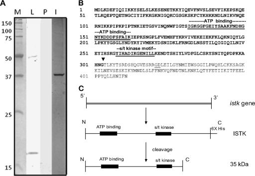
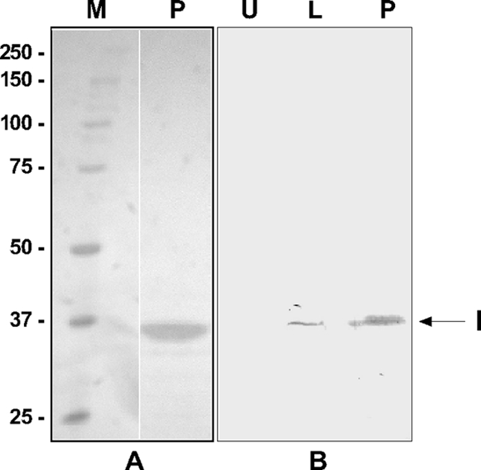
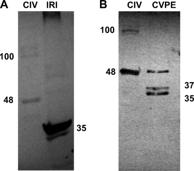
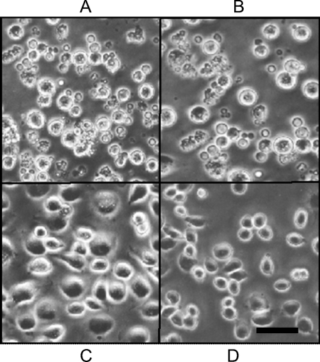
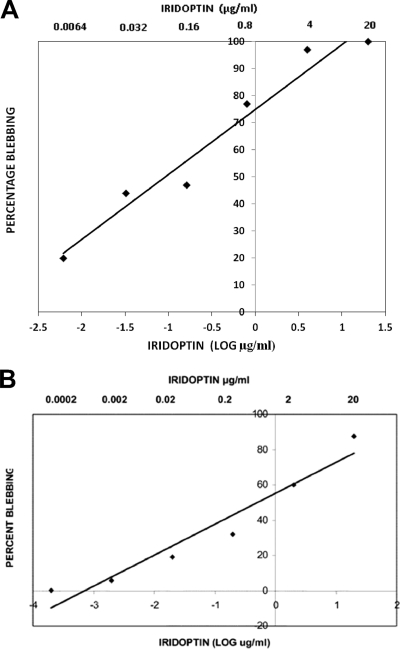
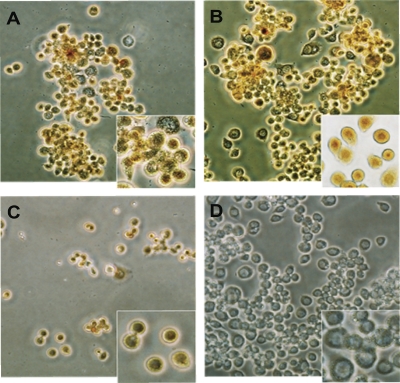
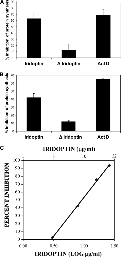
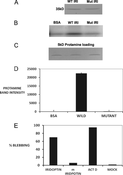
Apoptosis and inhibition of host gene expression are often associated with virus infections. Many viral polypeptides modulate apoptosis by direct interaction with highly conserved apoptotic pathways. Some viruses induce apoptosis during late stages of the infection cycle, while others inhibit apoptosis to facilitate replication or maintain persistent infection. In previous work, we showed that Chilo iridescent virus (CIV) or CIV virion protein extract induces apoptosis in spruce budworm and cotton boll weevil cell cultures. Here, we characterize the product of a CIV gene (iridovirus serine/threonine kinase; istk) with signature sequences for S/T kinase and ATP binding. ISTK appears to belong to the superfamily, vaccinia-related kinases (VRKs). The istk gene was expressed in Pichia pastoris vectors. Purified ISTK (48 kDa) exhibited S/T kinase activity. Treatment with ISTK induced apoptosis in budworm cells. A 35-kDa cleavage product of ISTK retaining key signature sequences was identified during purification. Pichia-expressed 35-kDa polypeptide, designated iridoptin, induced apoptosis and inhibition of host protein synthesis in budworm and boll weevil cells. A mutation in the ATP-binding site eliminated both kinase and apoptosis activity of iridoptin, suggesting that kinase activity is essential for induction of apoptosis. Analysis with custom antibody confirmed that ISTK is a structural component of CIV particles. This is the first demonstration of a viral kinase inducing apoptosis in any virus-host system and the first identification of a factor inducing apoptosis or host protein shutoff for the family Iridoviridae.


Figures

Fig. 1.
Fig. 1.
(A) Western analysis of ISTK with anti-His6 antibody. ISTK was expressed in P. pastoris and analyzed as described in Materials and Methods. Lane M, molecular mass markers (kDa); lane L, Pichia lysate showing ISTK (48 kDa) and a 17-kDa polypeptide; lane P, purified eluate from nickel-affinity column after imidazole treatment; lane I, silver stain of the eluate in lane P showing a 35-kDa band. (B) Amino acid sequence of ISTK. The motifs for ATP binding and S/T kinase are underlined. The ISTK cleavage site resulting in the 35-kDa product (bold type) during downstream processing of ISTK is shown (▾). (C) Downstream processing of ISTK and derivation of active 35-kDa polypeptide from the ISTK gene product.
Fig. 2.
Fig. 2.
SDS-PAGE analysis of His-tagged iridoptin expressed in the Pichia system and purified on Ni-affinity columns. (A) Silver stain analysis. Lane M, molecular mass (kDa) markers; lane P, iridoptin purified on ProBond columns. Note the 35-kDa band. (B) Western analysis with anti-His6 antibody. Lane U, uninduced yeast lysate; lane L, induced lysate showing 35-kDa iridoptin band (I); lane P, iridoptin purified on ProBond affinity columns.
Fig. 3.
Fig. 3.
Immunoblot analysis of CIV polypeptides with custom antibody to the synthetic peptide I64-C77 from the iridoptin sequence. (A) CIV, 5 μg of purified CIV; IRI, 4 μg of iridoptin. (B) CIV, 6 μg of purified CIV; CVPE, 10 μg of viral protein extract. The numbers represent molecular mass in kDa. See Materials and Methods for details.
Fig. 4.
Fig. 4.
Iridoptin induces apoptotic blebbing in spruce budworm (CF) cells. Cells in Nunc 60-well trays (5.6 × 10³ cells per well in 7 μl of medium) were treated with iridoptin (10 μg/ml) (A), actinomycin D (4 μg/ml) (B), heat-inactivated iridoptin (10 μg/ml) (C), or RBSS (D). The cells were then incubated at 28°C for 24 h and observed for blebbing by phase-contrast microscopy. Scale bar, 10 μm.
Fig. 5.
Fig. 5.
Dose-response analysis of iridoptin-induced apoptosis in budworm and boll weevil cells. (A) Budworm cells (CF) were treated with dilutions of iridoptin starting at 20 μg/ml (final concentration) and incubated at 28°C for 24 h. Cells were observed for apoptotic blebs with a phase-contrast microscope. Percent blebbing was determined from approximately 250 cells per field. Linear regression line and R2 value were generated using Microsoft Excel 2003. The linear regression line was generated with the following equation: y = 24x + 75, where x is the log of final iridoptin concentration in μg/ml and y is the percentage of cell population with blebs. The R2 value for the fitted line was 0.95. The highest dilution of toxin-produced blebbing in 50% of the cell population was approximately 0.1 μg/ml. (B) Boll weevil (AG) cells. The line was generated by the following equation: y = 17.5x + 55, where x is the log of final iridoptin concentration in μg/ml and y is the percentage of cell population with blebs. The R2 value for the fitted line was 0.94. The final concentration of iridoptin required to induce blebbing in 50% of the cell population was 0.5 μg/ml. Assays for both cell lines were performed in triplicate.
Fig. 6.
Fig. 6.
Confirmation of iridoptin-induced apoptosis in boll weevil (AG) cells by TUNEL assay. AG cells were grown on glass slides in CellStar six-well plates (5.6 × 10³ cells per well in 2.5 ml of medium). Cells were treated as described in Materials and Methods and viewed by phase-contrast microscopy. Brown staining of the nuclei indicated positive DAB signal. Assays were performed in duplicate. Treatments were as follows: 10 μg/ml iridoptin (A), 20 μg/ml iridoptin (B), 4 μg/ml actinomycin D (C), and mock treatment (D). Magnification, ×200. Insets show detail of nuclear DAB signal (magnification, ×400).
Fig. 7.
Fig. 7.
Iridoptin induces inhibition of protein synthesis in insect cells. Cells were pulsed with [35S]methionine 3 h after treatment with iridoptin (7 μg/ml), actinomycin D (ActD; 4 μg/ml), or heat-inactivated iridoptin (ΔIridoptin; 7 μg/ml). Polypeptides were separated on 10% SDS-polyacrylamide gels, and radioactive protein was visualized with a Typhoon 9410 (Molecular Dynamics) phosphorimager. Inhibition values are percentages of transmittance against mock lanes. (A) Budworm (CF) cells. (B) Boll weevil (AG) cells. (C) Dose-response analysis of iridoptin-induced host shutoff in boll weevil (AG) cells. A linear regression line was generated using Microsoft Excel 2003 and the following equation: y = 98.4x − 44.9, where x is the log of final iridoptin concentration in μg/ml and y is percent inhibition of host protein synthesis. The R2 value for the fitted line was 0.99. The final concentrations of iridoptin required to inhibit 50% and 90% host protein synthesis were 10 μg/ml and 23 μg/ml, respectively. Data are means of triplicate determinations.
Fig. 8.
Fig. 8.
Kinase activity and apoptosis induction by wild-type and mutant iridoptin. (A) Wild-type (WT) and mutant (Mut) iridoptin were separately purified from induced Pichia lysates using nickel affinity columns (ProBond), separated by SDS-PAGE, and stained with Coomassie blue. A single 35-kDa band was observed for each preparation. (B) Kinase activity. Assays were carried out as described in Materials and Methods. [γ-32P]ATP was used as the label, and protamine (5 kDa) was used as the substrate. (C) Substrate loading controls. (D) Band intensity mean integrated pixel density units of [32P]protamine substrate in plate B quantified with NIH ImageJ software. (E) CF cells treated with 9 μg/ml of iridoptin or kinase-dead mutant (m; K153Q) iridoptin and observed for apoptotic blebbing at 24 h. ActD, actinomycin D; Mock, RBSS. Percentages are means of three fields each comprising 150 cells.

Similar articles

Cited by

References

    1. Bablanian R., Banerjee A. K. 1986. Poly(riboadenylic acid) preferentially inhibits in vitro translation of cellular mRNAs compared with vaccinia virus mRNAs: possible role in vaccinia virus cytopathology. Proc. Natl. Acad. Sci. U. S. A. 83:1290–1294 - PMC - PubMed
    1. Banham A. H., Leader D. P., Smith G. L. 1993. Phosphorylation of ribosomal proteins by the vaccinia virus B1R protein kinase. FEBS Lett. 321:27–31 - PubMed
    1. Bilimoria S. L. March 2001. Use of viral proteins for controlling the cotton boll weevil and other insect pests. U.S. patent 6200561
    1. Bilimoria S. L., Sohi S. S. 1977. Development of an attached strain from a continuous insect cell line. In Vitro 13:461–466 - PubMed
    1. Bromenshenk J. J., et al. 2010. Iridovirus and microsporidian linked to honey bee colony decline. PLoS One 5:e13181. - PMC - PubMed

Publication types

MeSH terms

LinkOut - more resources