. 2011 Dec;85(24):13278-89.
doi: 10.1128/JVI.05583-11. Epub 2011 Oct 12.

Characterization of the pleiotropic effects of the genotype G-specific 36-nucleotide insertion in the context of other hepatitis B virus genotypes

Affiliations

Characterization of the pleiotropic effects of the genotype G-specific 36-nucleotide insertion in the context of other hepatitis B virus genotypes

Danielle Gutelius et al. J Virol. 2011 Dec.

Abstract

The pregenomic RNA (pgRNA) of hepatitis B virus (HBV) serves as the messenger for both core and P proteins, with the downstream P gene translated by ribosomal leaky scanning. HBV replication begins with packaging of the pgRNA and P protein into core protein particles, followed by conversion of RNA into DNA. Genotype G has a low replication capacity due to a low pgRNA level. It has a 36-nucleotide (nt) insertion in the 5' end of the core gene, adding 12 residues to the core protein. The insertion is needed to maintain efficient core protein expression and genome replication but causes inefficient virion secretion yet high maturity of virion DNA. In the present study, we confirmed that the 36-nt insertion had similar effects on core protein expression and virion secretion when it was introduced into genotype A and D clones but no impact on virion genome maturity. Surprisingly, the insertion impaired genome replication in both genotypes. Transcomplementation assays suggest that increased efficiency of core protein translation diminishes ribosomal scanning toward the downstream P gene. Indeed, mutating the core gene Kozak sequence restored core protein to lower levels but increased replication of the insertion mutant. Similar mutations impaired replication in genotype G. On the other hand, replacement of the core promoter sequence of genotype G with genotype A sequence increased pgRNA transcription and genome replication, implicating this region in the low replication capacity of genotype G. Why the 36-nt insertion is present in genotype G but absent in other genotypes is discussed.


Figures

Fig. 1.
Fig. 1.
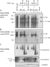
The 36-nt insertion impairs DNA replication in non-G genotypes. The insertion was introduced into three clones each of genotype A and genotype D. (A) Southern blot analysis of intracellular, core particle-associated HBV DNA. Lane M, the 3.2-kb and 1.7/1.5-kb HBV DNA size markers. RC, relaxed circular; PDS, partially double stranded; SS, single stranded. (B) Southern blot analysis of extracellular, virion-associated DNA following immunoprecipitation (IP-Southern). (C) Simultaneous analysis of intracellular core particles (upper panel) and core protein (middle panel). Core particles from cell lysate were separated by electrophoresis through nondenaturing native agarose gel and detected by a rabbit polyclonal anti-core antibody (Dako). Note retarded migration of core particles of clone 4B and its insertion mutant 4B+36 (C, lanes 3 and 4). Core protein from the same lysate was immunoprecipitated using a polyclonal rabbit antibody followed by Western blot analysis using the monoclonal antibody 14Ell (Abcam) (IP-Western). The 12-aa insertion retarded core protein migration in all insertion mutants. Western blot analysis of GAPDH from an aliquot of cell lysate served as a loading control (lower panel). (D) Northern blot analysis of HBV RNA transcripts in genotype A clones. The 3.5-kb pc/pgRNAs and subgenomic RNAs of 2.1 and 0.7 kb are indicated. (E) Ethidium bromide staining of the RNA gel prior to Northern blot analysis. The 28S and 18S bands are indicated and serve as loading controls.
Fig. 2.
Fig. 2.
The ε mutant of clone 4B+36, rather than its core/P mutant, is associated with diminished genome replication in the transcomplementation assay. The core/P mutant of 4B or 4B+36 was cotransfected with the ε mutant of 4B or 4B+36 at the indicated ratios. The core/P mutant served as the pgRNA for encapsidation and conversion to DNA while the ε mutant drove the expression of core and P proteins. Cells transfected with 1 μg of each mutant alone served as negative controls (lanes 1 to 4), while cells transfected with 1 μg of the parental clone 4B or 4B+36 served as positive controls (lanes 5 and 6). (A) Southern blot analysis of HBV DNA from intracellular core particles. (B) IP-Southern blot analysis of DNA from secreted virions. (C) IP-Western blot analysis of intracellular core protein. GAPDH was detected from an aliquot of cell lysate as a loading control.
Fig. 3.
Fig. 3.
Comparison of core protein expression constructs driven by the CMV promoter for their ability to reconstitute genome replication and virion secretion in core mutants. A core mutant of 4B or 4B+36 was transfected alone or cotransfected with each of the four CMV-core constructs (4B, 4B+36, G1, and G1−36) at 1 μg each. Cells transfected with 1 μg of the 4B or 4B+36 parental clone served as positive controls. (A) Southern blot analysis of intracellular HBV DNA. (B) IP-Southern blot analysis of virion-associated DNA. (C) Intracellular core particles separated by native agarose gel (upper panel) and core protein detected by IP-Western blot analysis (middle panel). GAPDH from the same cell lysate served as a loading control (lower panel).
Fig. 4.
Fig. 4.
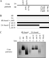
The amino acid substitutions at the N terminus of the 4B core protein are responsible for retarded core particle mobility. (A) Mutations in the core protein of genotype A clones 2A (top) and 4B (bottom). (B) Schematic diagram of chimeric constructs generated between clones 4B (filled box) and 2A (open box). (C) Mobility of intracellular core particles detected by native agarose gel electrophoresis.
Fig. 5.
Fig. 5.
The ε/P mutant, rather than the core mutant, determines genome replication capacity in the transcomplementation assay. The ε/P mutant of 4B or 4B+36 was cotransfected with the core mutant of 4B or 4B+36 at the indicated ratios. The ε/P mutant drove expression of the core protein while the core mutant served as the pgRNA for encapsidation and drove P protein expression. Cells transfected with 1 μg of the parental clone 4B or 4B+36 served as positive controls (lanes 1 and 2). (A) Southern blot analysis of HBV DNA from intracellular core particles. (B) Direct Western blot analysis of intracellular core protein using the polyclonal anti-core antibody (Dako) (upper panel). Please notice a nonspecific band comigrating with the core protein containing the 12-aa insertion. GAPDH was detected from the same blot following stripping (lower panel).
Fig. 6.
Fig. 6.
Effect of reducing core protein expression via Kozak sequence mutations on replication of genotype D (α1+36) and genotype G (G1). (A and E) Mutations made in the core gene Kozak sequence. Mutations are indicated in boldface and reduced font size. The initiation codon is underlined. (B and F) Southern blot analysis of intracellular replicative HBV DNA. (C and G) IP-Southern blot analysis of virion-associated HBV DNA. (D and H) IP-Western blot analysis of intracellular core protein (upper panel) and GAPDH loading control (lower panel). Densitometry was performed using ImageJ software. Quantification of core protein signal intensity is indicated as a percentage of the α1+36 or G1 parental value, which was set at 100. All values were normalized to the GAPDH control.
Fig. 7.
Fig. 7.
Effect of knocking down an internal core gene AUG codon on the replication capacity of HBVα1 and its insertion mutant (A to C) and the ability of the mutant core protein to rescue genome replication and virion secretion in the core mutant (D to F). C2KO mutants have the C2 AUG converted to AUC, which results in the M93I amino acid change in the core protein. (A and D) Southern blot analysis of intracellular HBV DNA. (B and E) IP-Southern blot analysis of virion-associated DNA. (C and F) Native agarose gel followed by Western blot analysis for core particle detection (F, top panel). IP-Western blot analysis of intracellular core protein with GAPDH serving as the loading control. For panels D to F, 1 μg of the 4B core dimer was cotransfected with 1 μg of the parental or M93I mutant CMV-core construct of α1+36.
Fig. 8.
Fig. 8.
Rescue of the replication capacity of G1 and the G1−36 mutant by replacement of the core promoter sequence. Nucleotides 1606 to 1766 of both clone G1 and G1−36 were replaced with 4B sequence to generate the M1 mutants. (A) Northern blot analysis of HBV RNA transcripts. The 3.5-kb pc/pgRNAs and subgenomic RNAs of 2.4, 2.1, and 0.7 kb are indicated. (B) Ethidium bromide staining of the RNA gel prior to Northern blot analysis. (C) Western blot analysis of intracellular core protein (upper panel) and GAPDH (lower panel). (D) Southern blot analysis of intracellular replicative DNA. (E) IP-Southern blot analysis of extracellular virion-associated DNA.
Fig. 9.
Fig. 9.
Characterization of a series of G1−36 mutants with exchange of core promoter (CP) sequence. (A) Schematic diagram of three chimeric constructs generated between clone 4B of genotype A (filled box) and G1−36 of genotype G (open box). (B) Additional CP mutants of G1−36 (M4 to M10) with partial replacement with 4B or wild-type genotype G sequence. Clone G1 has a single nucleotide deletion (nt 1721), as indicated by the minus sign. The Sp1-1 and Sp1-2 binding sites are indicated. (C) Southern blot analysis of intracellular DNA replication. (D) IP-Southern blot analysis for detection of secreted virions. (E) Detection of intracellular core particles, core protein, and GAPDH.

Similar articles

Cited by

References

    1. Bang G., et al. 2005. Effect of mutating the two cysteines required for HBe antigenicity on hepatitis B virus DNA replication and virion secretion. Virology 332:216–224 - PubMed
    1. Bhat R. A., Ulrich P. P., Vyas G. N. 1990. Molecular characterization of a new variant of hepatitis B virus in a persistently infected homosexual man. Hepatology 11:271–276 - PubMed
    1. Blum H. E., et al. 1992. Hepatitis B virus X protein is not central to the viral life cycle in vitro. J. Virol. 66:1223–1227 - PMC - PubMed
    1. Chen A., Kao Y. F., Brown C. M. 2005. Translation of the first upstream ORF in the hepatitis B virus pregenomic RNA modulates translation at the core and polymerase initiation codons. Nucleic Acids Res. 33:1169–1181 - PMC - PubMed
    1. Cotelesage J. J., et al. 2011. Hepatitis B virus genotype G forms core-like particles with unique structural properties. J. Viral Hepat. 18:443–448 - PMC - PubMed

Publication types

Substances