. 2009 Sep;1(2):298-316.
doi: 10.3390/v1020298. Epub 2009 Sep 10.

Viroid pathogenicity: one process, many faces

Affiliations

Viroid pathogenicity: one process, many faces

Robert A Owens et al. Viruses. 2009 Sep.

Abstract

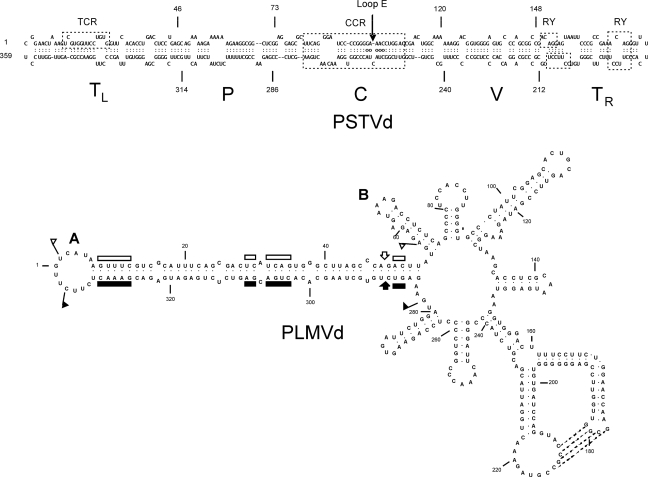
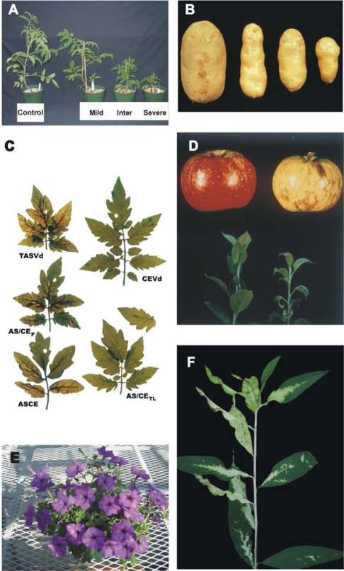
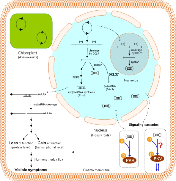
Despite the non-coding nature of their small RNA genomes, the visible symptoms of viroid infection resemble those associated with many plant virus diseases. Recent evidence indicates that viroid-derived small RNAs acting through host RNA silencing pathways play a key role in viroid pathogenicity. Host responses to viroid infection are complex, involving signaling cascades containing host-encoded protein kinases and crosstalk between hormonal and defense-signaling pathways. Studies of viroid-host interaction in the context of entire biochemical or developmental pathways are just beginning, and many working hypotheses have yet to be critically tested.

Keywords: RNA silencing; disease induction; viroid pathogenicity.


Figures

Figure 1.
Figure 1.
Secondary structures of PSTVd and Peach latent mosaic viroid (PLMVd). (A) The rod-like secondary structure of PSTVd (intermediate strain) showing the five domains characteristic of members of the family Pospiviroidae: the Terminal Left (TL), Pathogenicity (P), Central (C), Variable (V), and Terminal Right (TR). The Central Conserved Region (CCR) is located within the C domain and contains a UV-sensitive loop E motif with non-canonical base-pairs (denoted by circles). The TL domain of posiviroids contains either a Terminal Conserved Region (TCR) or Terminal Conserved Hairpin (location not shown). The TR domain may contain 1–2 copies of a protein-binding RY motif [16]. (B) The branched secondary structure of PLMVd, a member of the ribozyme-containing family Avsunviroidae. Boundaries of the plus and minus strand self-cleavage domains are indicated by flags, nucleotides conserved in most natural hammerhead structures by bars, and the self-cleavage sites by arrows. Filled and open symbols refer to plus and minus polarities, respectively. Nucleotides involved in a pseudoknot supported by chemical probing are indicated by broken lines. Co-variation analysis suggests that a second pseudoknot may exist between loops A (location of the short insertion responsible for peach calico) and B.
Figure 2.
Figure 2.
Symptoms associated with viroid infection. (A,C) Infection of sensitive tomato cultivars with PSTVd or related viroids like CEVd and TASVd leads to stunting, epinasty (a downward curling of the leaves), and veinal necrosis. Note the differences in symptom severity associated with different strains of PSTVd (A) or different viroids (C). (B) Symptoms of PSTVd in its natural host (potato); the control tuber on the left is from a healthy plant. (D) Fruit from viroid-infected woody hosts like apple or plum may exhibit abnormal pigmentation; i.e., “color break”. Left, healthy apple; right, Apple scar skin viroid (ASSVd)-infected apple. (F) Infection of peach by certain variants of PLMVd leads to extreme chlorosis and loss of chlorophyll from large portions of the leaves (courtesy of Francesco Di Serio). (E) Many viroid-infected plants (especially herbaceous ornamentals like this Tomato chlorotic dwarf viroid (TCDVd)-infected petunia) may show no visible symptoms. Vegetative propagation of latently infected plant material dramatically increases the number of viroid-infected plants, thereby increasing the opportunity for accidental transfer (i.e., “escape”) to other sensitive species growing nearby.
Figure 3.
Figure 3.
Schematic overview of viroid pathogenicity illustrating both direct and indirect interaction between the viroid genome and host cell. As discussed by Gomez et al. [69], RNA silencing mediated by (vd)ta-siRNAs appears likely to play a major role in disease induction by viroids like PSTVd or HSVd that replicate in the nucleus. The resulting cleavage of host mRNAs could lead to either loss or gain of function at protein level. PSTVd (and presumably other viroid RNAs) also activates at least two protein kinases, one of which (PKV) may be associated with the plasma membrane. Activation of the signaling cascades containing these kinases would then lead to perturbations in plant defense and hormone signaling pathways. Very little is known about siRNA synthesis associated with viroid replication in the chloroplast. Viroid replication in both the nucleus and cytoplasm proceeds via a rolling circle mechanism (indicated by open circles with opposing arrows). For viroids like PSTVd that replicate in the nucleus, various stages of replication appear to be localized in either the nucleoplasm or the nucleolus [75].

Similar articles

Cited by

References

    1. Diener TO. Potato spindle tuber “virus” IV. A replicating, low molecular weight RNA. Virology. 1971;45:411–428. - PubMed
    1. Gross HJ, Domdey H, Lossow C, Jank P, Raba M, Alberty H, Sänger H-L. Nucleotide sequence and secondary structure of potato spindle tuber viroid. Nature. 1978;273:203–208. - PubMed
    1. Dickson E, Robertson HD, Niblett CL, Horst RK, Zaitlin M. Minor differences between nucleotide sequences of mild and severe strains of potato spindle tuber viroid. Nature. 1979;277:60–62.
    1. Keese P, Symons RH. Domains in viroids: Evidence of intermolecular RNA rearrangements and their contribution to viroid evolution. Proc Natl Acad Sci USA. 1985;82:4582–4586. - PMC - PubMed
    1. Schnölzer M, Haas B, Ramm K, Hofmann H, Sänger H-L. Correlation between structure and pathogenicity of potato spindle tuber viroid (PSTV) EMBO J. 1985;4:2181–2190. - PMC - PubMed

LinkOut - more resources