. 2009 Sep;1(2):317-34.
doi: 10.3390/v1020317. Epub 2009 Sep 14.

Viroid replication: rolling-circles, enzymes and ribozymes

Affiliations

Viroid replication: rolling-circles, enzymes and ribozymes

Ricardo Flores et al. Viruses. 2009 Sep.

Abstract

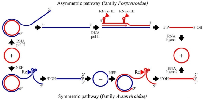
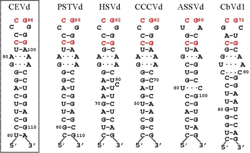
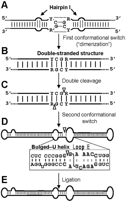
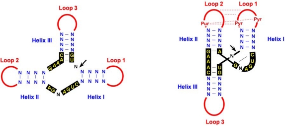
Viroids, due to their small size and lack of protein-coding capacity, must rely essentially on their hosts for replication. Intriguingly, viroids have evolved the ability to replicate in two cellular organella, the nucleus (family Pospiviroidae) and the chloroplast (family Avsunviroidae). Viroid replication proceeds through an RNA-based rolling-circle mechanism with three steps that, with some variations, operate in both polarity strands: i) synthesis of longer-than-unit strands catalyzed by either the nuclear RNA polymerase II or a nuclear-encoded chloroplastic RNA polymerase, in both instances redirected to transcribe RNA templates, ii) cleavage to unit-length, which in the family Avsunviroidae is mediated by hammerhead ribozymes embedded in both polarity strands, while in the family Pospiviroidae the oligomeric RNAs provide the proper conformation but not the catalytic activity, and iii) circularization. The host RNA polymerases, most likely assisted by additional host proteins, start transcription from specific sites, thus implying the existence of viroid promoters. Cleavage and ligation in the family Pospiviroidae is probably catalyzed by an RNase III-like enzyme and an RNA ligase able to circularize the resulting 5' and 3' termini. Whether a chloroplastic RNA ligase mediates circularization in the family Avsunviroidae, or this reaction is autocatalytic, remains an open issue.

Keywords: catalytic RNAs; hammerhead ribozymes; viroids.


Figures

Figure 1.
Figure 1.
Rolling-circle replication mechanism with the two alternative pathways followed by members of the family Pospivirodae (asymmetric) and Avsunviroidae (symmetric) [15,16,46]. Red and blue lines refer to (+) and (−) strands, respectively. Arrowheads mark the cleavage sites of a host RNase III or a hammerhead ribozymes (Rz), and the resulting terminal groups are indicated. Elongation of RNA strands is catalyzed by the nuclear RNA polymerase II (RNA pol II) or the nuclear-encoded chloroplastic RNA polymerase (NEP).
Figure 2.
Figure 2.
Hairpin I structures of the five type species of the family Pospiviroidae. This structural element is formed by the upper CCR strand and flanking nucleotides of the type members of the five genera composing the family Pospiviroidae: PSTVd, HSVd, CCCVd (Coconut cadang-cadang viroid), ASSVd, and CbVd1 (Coleus blumei viroid 1) [25,26]. Red fonts indicate conserved nucleotides in structurally similar positions. Continuous and broken lines represent Watson-Crick and non-canonical base pairs, respectively [28]. Notice that the variability preserves the overall structure of hairpin I, including the terminal palindromic tetraloop, the adjacent 3-bp stem, and the long stem. Left inset, hairpin I of the CEVd variant used to transform A. thaliana [26]; notice two covariations with respect to PSTVd at the basis of the long stem. Reprinted with permission from [26].
Figure 3.
Figure 3.
Model for processing in vivo of the oligomeric (+) replicative intermediates of the family Pospiviroidae that involves a kissing loop interaction between the palindromic tetraloops of two consecutive hairpin I motifs (A), with their stems forming subsequently a longer interstrand duplex (B). This double-stranded structure is the substrate for cleavage at specific positions in both strands (C). Following a second conformational switch the resulting unit-length strands adopt the extended rod-like structure with loop E (in outlined fonts) and the adjacent bulged-U helix (D), which is the substrate for ligation (E). R and Y refer to purines and pyrimidines, respectively, the S-shaped line denotes the UV-induced cross-link, and white arrowheads mark the cleavage sites in the double-stranded structure and the ligation site in the extended conformation. Reprinted with permission from [26].
Figure 4.
Figure 4.
Hammerhead structures. (A) Schematic representation of a typical hammerhead structure as originally proposed [45]. Residues strictly or highly conserved in natural hammerhead structures are in yellow on a black background. Arrow marks the self-cleavage site, N indicates any residue and H any residue except G, and continuous and broken lines denote Watson-Crick and non-canonical base pairs, respectively. (B) Hammerhead structure represented according to X-ray crystallography and NMR data [–62]. The proposed tertiary interaction between loops 1 and 2, which facilitates catalytic activity in vivo, is indicated with red broken lines. Pyr and Pur refer to pyrimidine and purine, respectively.

Similar articles

Cited by

References

    1. Gross HJ, Domdey H, Lossow C, Jank P, Raba M, Alberty H, Sänger HL. Nucleotide sequence and secondary structure of potato spindle tuber viroid. Nature. 1978;273:203–208. - PubMed
    1. Diener TO. Discovering viroids—a personal perspective. Nat Rev Microbiol. 2003;1:75–80. - PubMed
    1. Tabler M, Tsagris M. Viroids: petite RNA pathogens with distinguished talents. Trends Plant Sci. 2004;9:339–348. - PubMed
    1. Flores R, Hernández C, Martínez de Alba E, Daròs JA, Di Serio F. Viroids and viroid-host interactions. Annu Rev Phytopathol. 2005;43:117–139. - PubMed
    1. Daròs JA, Elena SF, Flores R. Viroids: an Ariadne’s thread into the RNA labyrinth. EMBO Rep. 2006;7:593–598. - PMC - PubMed

LinkOut - more resources