. 2009 Dec;1(3):895-919.
doi: 10.3390/v1030895. Epub 2009 Nov 10.

Defective Interfering RNAs: Foes of Viruses and Friends of Virologists

Affiliations

Defective Interfering RNAs: Foes of Viruses and Friends of Virologists

Kunj B Pathak et al. Viruses. 2009 Dec.

Abstract

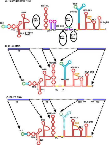
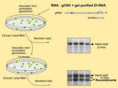
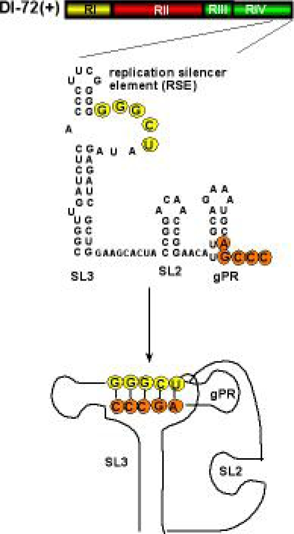
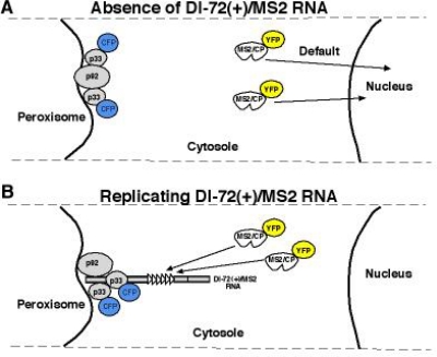
Defective interfering (DI) RNAs are subviral RNAs produced during multiplication of RNA viruses by the error-prone viral replicase. DI-RNAs are parasitic RNAs that are derived from and associated with the parent virus, taking advantage of viral-coded protein factors for their multiplication. Recent advances in the field of DI RNA biology has led to a greater understanding about generation and evolution of DI-RNAs as well as the mechanism of symptom attenuation. Moreover, DI-RNAs are versatile tools in the hands of virologists and are used as less complex surrogate templates to understand the biology of their helper viruses. The ease of their genetic manipulation has resulted in rapid discoveries on cis-acting RNA replication elements required for replication and recombination. DI-RNAs have been further exploited to discover host factors that modulate Tomato bushy stunt virus replication, as well as viral RNA recombination. This review discusses the current models on generation and evolution of DI-RNAs, the roles of viral and host factors in DI-RNA replication, and the mechanisms of disease attenuation.

Keywords: RNA structure; RNA virus; host factors; recombination; replication.


Figures

Figure 1.
Figure 1.
Genome and structural organization of TBSV genomic RNA and the prototypical DI-73 and DI-72 RNAs. (A) A cartoon showing structural details of the ∼4,800 nt TBSV genome (not to scale). The p33, p92pol, p41 and the overlapping p19/p22 ORFs are depicted as black ovals and labeled accordingly. Note that p92pol overlaps with p33, sharing the same initiation codon. Sequences playing role(s) in translation, genome replication and sgRNA transcription are shown in turquoise blue, red and purple, respectively. Sequences involved in RNA-RNA interactions are shown in matching colors. Note that translation requires SL3-SLB interaction, UL-DL and RSE-gPR interactions are required for replicase assembly and AS1-RS1, AS2-RS2 and DE-CE interactions are crucial for sgRNA synthesis. (Abbreviations used are DSD: downstream domain; TSD: T-shape domain: RSE: replication silencer element; AS: activator sequence; RS: receptor sequence; CE: core element; DE: distal element; UL: upstream linker; DL: downstream linker; CITE: cap independent translation enhancer; SL: stem loop) [104] (B) Structure of the ∼800 nt DI-73 carrying three noncontiguous segments of the genomic RNA. Generation of DI-73 preserves critical replication elements (red) and the 3′CITE. The blue bars and dotted arrows depict the segments corresponding to genomic RNA. (C) Note that the other prototypical DI of ∼620 nt, named DI-72 RNA, has an additional deletion of the 3′CITE.
Figure 2.
Figure 2.
Experimental scheme to test DI-RNA evolution in plant protoplasts. Generation of DI-RNA recombinants are tested via using the total RNA extract from the first protoplast samples for electroporation of the second batch of protoplasts (1st passage). The images on the right show Northern blot analysis of the total RNA extracts used to detect the original DI-RNA and the recombinants [41,42].
Figure 3.
Figure 3.
The predicted secondary and tertiary structure of the 3′ UTR in DI-72(+) RNA. A 5 nt base-pairing between RSE in the internal loop sequence of SL3 and gPR stabilizes the tertiary structure. This interaction is critical for the assembly of the functional TBSV replicase [95,112].
Figure 4.
Figure 4.
Different subcellular localization of plus- and minus-stranded DI-72 RNA during replication in yeast. (A) Schematic presentation of subcellular localization of MS2/CP-YFP and CFP-p33 in yeast in the absence of viral RNA and (B) in the presence of DI-72(+)/MS2. Specific interaction between the MS2 CP and the MS2 CP recognition hairpin (present in six copies in DI-72(+)/MS2) should result in re-localization of MS2 CP as shown. (C) Co-localization of CFP-tagged p33 and the YFP-tagged MS2/CP bound to the DI-72(+)/MS2 RNA in yeast cells using epifluorescence microscopy. Note that DI-72(–)/MS2 RNA (bottom panel) contains the six copies of MS2 CP recognition hairpins in complementary orientation. Therefore, MS2/CP-YFP could only bind to the negative-stranded DI RNA, which is generated during the replication of DI-72(–)/MS2 RNA.
Figure 4.
Figure 4.
Different subcellular localization of plus- and minus-stranded DI-72 RNA during replication in yeast. (A) Schematic presentation of subcellular localization of MS2/CP-YFP and CFP-p33 in yeast in the absence of viral RNA and (B) in the presence of DI-72(+)/MS2. Specific interaction between the MS2 CP and the MS2 CP recognition hairpin (present in six copies in DI-72(+)/MS2) should result in re-localization of MS2 CP as shown. (C) Co-localization of CFP-tagged p33 and the YFP-tagged MS2/CP bound to the DI-72(+)/MS2 RNA in yeast cells using epifluorescence microscopy. Note that DI-72(–)/MS2 RNA (bottom panel) contains the six copies of MS2 CP recognition hairpins in complementary orientation. Therefore, MS2/CP-YFP could only bind to the negative-stranded DI RNA, which is generated during the replication of DI-72(–)/MS2 RNA.
Figure 5.
Figure 5.
Schematic presentation of the phosphorylation sites in the TBSV p33 replication protein. The phosphorylated aminoacids are shown in red, while the RNA binding site is indicated with blue letters. The negative charges of phosphorylated amino acids and the positive charges of arginines are shown. Possible interactions are depicted with dotted lines. Note that the RNA has also negative charge, resulting its release from p33 after phosphorylation of p33 as shown [109].

References

    1. Huang AS. Defective interfering viruses. Annu Rev Microbiol. 1973;27:101–117. - PubMed
    1. Simon AE, Roossinck MJ, Havelda Z. Plant virus satellite and defective interfering RNAs: new paradigms for a new century. Annu Rev Phytopathol. 2004;42:415–437. - PubMed
    1. Perrault J. Origin and replication of defective interfering particles. Curr Top Microbiol Immunol. 1981;93:151–207. - PubMed
    1. Qiu W, Scholthof KB. Defective interfering RNAs of a satellite virus. J Virol. 2001;75:5429–5432. - PMC - PubMed
    1. Luytjes W, Gerritsma H, Spaan WJ. Replication of synthetic defective interfering RNAs derived from coronavirus mouse hepatitis virus-A59. Virology. 1996;216:174–183. - PMC - PubMed

LinkOut - more resources