. 2009 Dec;1(3):1110-36.
doi: 10.3390/v1031110. Epub 2009 Dec 3.

HIV-1 Protease: Structural Perspectives on Drug Resistance

Affiliations

HIV-1 Protease: Structural Perspectives on Drug Resistance

Irene T Weber et al. Viruses. 2009 Dec.

Abstract

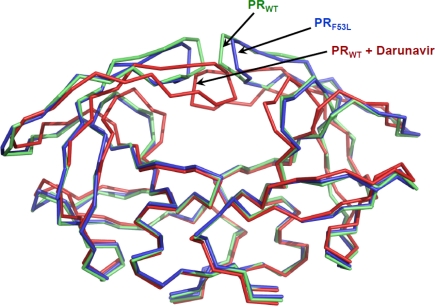
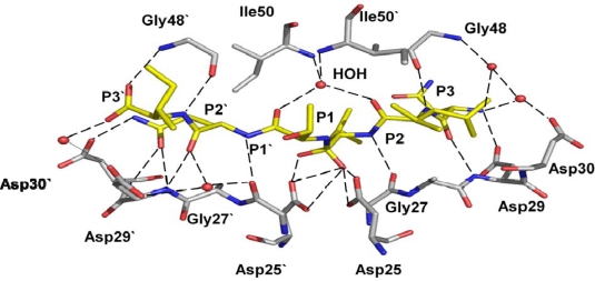
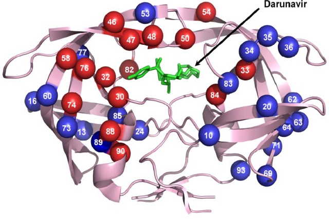
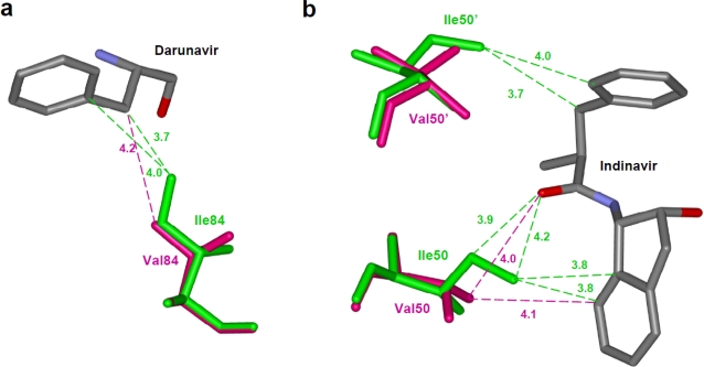
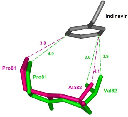
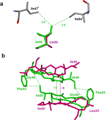
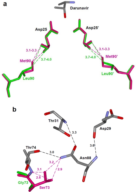
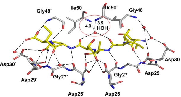
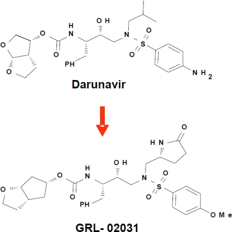
Antiviral inhibitors of HIV-1 protease are a notable success of structure-based drug design and have dramatically improved AIDS therapy. Analysis of the structures and activities of drug resistant protease variants has revealed novel molecular mechanisms of drug resistance and guided the design of tight-binding inhibitors for resistant variants. The plethora of structures reveals distinct molecular mechanisms associated with resistance: mutations that alter the protease interactions with inhibitors or substrates; mutations that alter dimer stability; and distal mutations that transmit changes to the active site. These insights will inform the continuing design of novel antiviral inhibitors targeting resistant strains of HIV.

Keywords: aspartic protease; darunavir; drug resistance; molecular mechanism; protease inhibitors.


Figures

Figure 1.
Figure 1.
Structures of HIV-1 protease dimer. Superposition of unliganded protease (PRWT in blue, PDB ID: 1HHP [29]), unliganded protease with F53L mutation (PRF53L in green, PDB ID: 2G69, [28]) and protease complex with darunavir (red, PDB ID: 2IEN, [30] inhibitor is removed for clarity). The unliganded structures exhibit opened flap conformation, while the protease flaps form the closed conformation with darunavir.
Figure 2.
Figure 2.
Hydrogen bond interactions of the peptide tetrahedral intermediate with HIV-1 protease (PDB ID: 3B7V [32]). The peptide intermediate is in yellow bonds and the protease in grey. Hydrogen bonds are indicated by broken lines. Interactions with the catalytic Asp25 and 25’ are omitted for clarity.
Figure 3.
Figure 3.
Sites of the resistance mutations on protease dimer. The protease dimer is in pink ribbons with darunavir in green sticks. Major and minor resistance mutations are colored as red and blue spheres, respectively. Mutations are distributed on both the monomers to increase visibility.
Figure 4.
Figure 4.
Resistance mutations showing loss of direct interactions with the inhibitor. (a) The I84V substitution results in loss of van der Waals contacts between residue 84 and darunavir. Wild type Ile84 (PDB ID: 2IEN) and mutant Val84 (PDB ID: 2IEO) are shown in green and magenta sticks, respectively. Only part of darunavir (grey bonds) is shown for clarity [30]. (b) Drug resistant mutation I50V is accompanied by loss of several interactions with indinavir [61]. Wild type Ile50 (PDB ID: 1SDT) and mutant Val (PDB ID: 2AVS) are shown as green and magenta sticks. Only the central portion of indinavir is shown in grey. The interatomic distances are given in Å.
Figure 5.
Figure 5.
The V82A mutation shows a shift in the main chain atoms of residues 81 and 82 that partially compensates for the loss of interactions due to substitution of a smaller side chain [64]. The wild type Val82 (PDB ID: 1SDT) and Ala mutant (PDB ID: 1SDV) are shown as green and magenta sticks, respectively.
Figure 6.
Figure 6.
(a)The I50V mutation at the tip of the flap results in loss of intersubunit interactions with Ile47’ and Ile84’ [61]. Ile50 (PDB ID: 1SDT) and Val50 (PDB ID: 2AVS) are represented as green and magenta sticks, respectively. (b) The F53L variant eliminates intersubunit hydrophobic interactions between residues 53 and Ile50’ [28]. This loss of interaction is accompanied by a wider separation of the flaps. The wild type (PDB ID: 1HHP) and F53L flaps (PDB ID: 2G69) are shown in green and magenta sticks. The separation between the flaps is indicated in Å.
Figure 7.
Figure 7.
(a) The longer methionine side chain of Met90 in the L90M variant forms shorter van der Waals interactions with the main chain of catalytic Asp25, unlike the Leu90 in the wild type protease [68]. The wild type (PDB ID: 2IEN) and mutant protease (PDB ID: 2F81) are shown in green and magenta colored sticks, respectively. Part of darunavir is shown colored by element type. (b) The protease variant with G73S (PDB ID: 2AVV) substitution forms new hydrogen bond interactions of Ser73 with Thr74 and Asn88 (magenta dashed lines) [61]. The hydrogen bond network of Thr74, Asn88, Asp29 and Thr31 propagates the effects to the active site cavity.
Figure 8.
Figure 8.
The variant I54V (PDB ID: 2B80) with peptide tetrahedral intermediate has lost the water mediated interactions with Ile50 and Ile50’, as indicated by red circle, in comparison to the wild type protease interactions in Figure 2 [32]. The hydrogen bond interactions between the protease and the intermediate are shown as broken lines.
Figure 9.
Figure 9.
Chemical structures of darunavir and the new antiviral inhibitor GRL-02031.

Similar articles

Cited by

References

    1. Weber IT, Zhang Y, Tozser J. HIV-1 Protease and AIDS Therapy. In: Lendeckel U, Hooper N, editors. Viral Proteases And Antiviral Protease Inhibitor Therapy. in press.
    1. Palella FJ, Jr, Delaney KM, Moorman AC, Loveless MO, Fuhrer J, Satten GA, Aschman DJ, Holmberg SD. Declining morbidity and mortality among patients with advanced human immunodeficiency virus infection. HIV Outpatient Study Investigators. N Engl J Med. 1998;338:853–860. - PubMed
    1. Hammer SM, Squires KE, Hughes MD, Grimes JM, Demeter LM, Currier JS, Eron JJ, Jr, Feinberg JE, Balfour HH, Jr, Deyton LR, Chodakewitz JA, Fischl MA. A controlled trial of two nucleoside analogues plus indinavir in persons with human immunodeficiency virus infection and CD4 cell counts of 200 per cubic millimeter or less. AIDS Clinical Trials Group 320 Study Team. N Engl J Med. 1997;337:725–733. - PubMed
    1. Sepkowitz KA. AIDS--the first 20 years. N Eng J Med. 2001;344:1764–1772. - PubMed
    1. Ghosh AK, Chapsal BD, Weber IT, Mitsuya H. Design of HIV protease inhibitors targeting protein backbone: an effective strategy for combating drug resistance. Acc Chem Res. 2008;41:78–86. - PubMed

LinkOut - more resources