. 2010 Dec;2(12):2618-48.
doi: 10.3390/v2122618. Epub 2010 Dec 3.

Jaagsiekte sheep retrovirus biology and oncogenesis

Affiliations

Jaagsiekte sheep retrovirus biology and oncogenesis

Andrew Hofacre et al. Viruses. 2010 Dec.

Abstract

Jaagsiekte sheep retrovirus (JSRV) is the causative agent of a lung cancer in sheep known as ovine pulmonary adenocarcinoma (OPA). The disease has been identified around the world in several breeds of sheep and goats, and JSRV infection typically has a serious impact on affected flocks. In addition, studies on OPA are an excellent model for human lung carcinogenesis. A unique feature of JSRV is that its envelope (Env) protein functions as an oncogene. The JSRV Env-induced transformation or oncogenesis has been studied in a variety of cell systems and in animal models. Moreover, JSRV studies have provided insights into retroviral genomic RNA export/expression mechanisms. JSRV encodes a trans-acting factor (Rej) within the env gene necessary for the synthesis of Gag protein from unspliced viral RNA. This review summarizes research pertaining to JSRV-induced pathogenesis, Env transformation, and other aspects of JSRV biology.

Keywords: Env; JSRV; Jaagsiekte sheep retrovirus; Rej; cancer; ovine pulmonary adenocarcinoma; transformation.


Figures

Figure 1
Figure 1
Genomic organization of JSRV. The JSRV genome is that of a betaretrovirus, possessing only the minimal number of genes required to carryout the retrovirus lifecycle; gag, pro, pol, and env. Uniquely, JSRV also has an additional open reading frame, orf-x, of unknown function. The boxes represent ORFs. LTR; long terminal repeats [2].
Figure 2
Figure 2
The structure of a typical retrovirus. All retroviruses carry two copies of their RNA genome (vRNA) and are composed of the viral structural proteins (SU, TM, MA, NC, and CA) and the viral enzymes (RT, PR, and IN).
Figure 3
Figure 3
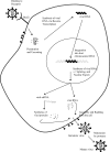
The retroviral life cycle. See text for details.
Figure 4
Figure 4
Clinical characteristics of ovine pulmonary adenocarcinoma. (A) OPA affected animals develop progressive respiratory distress, reflected by the accumulation of fluid within the respiratory tract. (B) Sheep lungs from an animal with OPA. The lungs display lesions (arrowed) characteristic of OPA as well as exudation of foamy lung fluid from the trachea. Adapted from [36].
Figure 5
Figure 5
Molecular clone of the exogenous JSRV21 isolate. pJSRV21 is a plasmid containing the full-length integrated proviral sequences of JSRV21. The JSRV 5’ LTR was replaced with the CMV immediate early promoter (pCMV2JS21) in order to drive robust expression of the JSRV21 sequences in a multitude of cell lines that normally do not display high transcription of the JSRV LTR [21]. pCMVJS21ΔGP is a derivative construct of pCMV2JS21 in which the reading frames for Gag/Pol have been removed and expresses only the JSRV Env. The env gene splice donor (sd) and splice acceptor (sa) sites are retained in plasmid ΔGP, necessary for efficient Env expression. Adapted from [43].
Figure 6
Figure 6
Transformation by the JSRV Env in NIH 3T3 fibroblasts. (A) The JSRV env expression plasmid (ΔGP) is driven by the constitutively active CMV promoter. (B) Demonstration of JSRV Env induced foci formation in NIH 3T3 cells transfected with ΔGP. Adapted from [43].
Figure 7
Figure 7
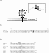
Schematic representation of the JSRV Env protein at the cell plasma membrane and sequence alignments of the Env cytoplasmic tail. (A) Hypothetical model of the JSRV Env (on a virion) with the JSRV receptor, Hyal2, and its likely positioning in the cellular membrane (insert). The JSRV Env is composed of two subunits, surface (SU) and transmembrane (TM). The cytoplasmic tail (CT) of the TM domain contains a YXXM motif, a putative PI3K/p85 binding site. (B) Alignment of the amino acid sequence of the TM region of the exogenous JSRV21 and the endogenous enJS56A1 and enJS5F16 clones. A dash (−) indicates lack of an amino acid. The amino acids of the putative PI3K docking site are in bold. (C) The putative PI3K docking site is conserved between various exJSRV isolates. Adapted from [75].
Figure 8
Figure 8
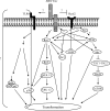
Signaling pathways involved in JSRV Env-induced cell transformation. Depending on the type of cell line studied, three main pathways have been shown to be activated in JSRV transformed cells; the PI3K/Akt pathway, the Hyal2-RON pathway, and the Ras-MEK-ERK pathway. Signaling through the TLR4-NFkB pathway is also depicted, since the JSRV Env interacts with TLR4, though activation of this pathway has yet to be formally shown. A fourth pathway; via signaling through Rac1, has recently been identified, but less is known about it [86]. Except for the Hyal2-RON pathway, how the JSRV Env engages the Ras-MEK-ERK or the PI3K/Akt pathways is not known. Possible interactions, or yet to be identified factors that may be involved in JSRV Env-mediated transformation, are indicated with question marks (?). Each pathway involved in JSRV Env-transformation leads to the activation of factors involved in cell survival, proliferation, apoptosis, and differentiation. See text for details.
Figure 9
Figure 9
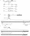
Schematic representation of the alternatively spliced transcripts within the JSRV env gene and the domains located within the Env signal peptide region. (A) Structure of doubly spliced env mRNAs with the env splice donor (sd) and splice acceptor (sa) nucleotides indicated. The locations of stop codons are indicated by asterisks. (B) Domain structures of the signal peptide of the JSRV Env. The JSRV Env signal peptide sequence is diagramed, and the locations of predicted sequence motifs are indicated by arrows under the diagram. (C) The putative translation products of the doubly spliced JSRV env transcripts. The locations of the alternative splice donor and splice acceptor sites are indicated, as well as the location of the stop codon (asterisks) within the second exons (shaded) [129].

References

    1. DeMartini JC, Bishop JV, Allen TE, Jassim FA, Sharp JM, de las Heras M, Voelker DR, Carlson JO. Jaagsiekte sheep retrovirus proviral clone JSRV(JS7), derived from the JS7 lung tumor cell line, induces ovine pulmonary carcinoma and is integrated into the surfactant protein A gene. J Virol. 2001;75:4239–4246. - PMC - PubMed
    1. Palmarini M, Sharp JM, de las Heras M, Fan H. Jaagsiekte sheep retrovirus is necessary and sufficient to induce a contagious lung cancer in sheep. J Virol. 1999;73:6964–6972. - PMC - PubMed
    1. Fan H, Palmarini M, DeMartini JC. Transformation and oncogenesis by jaagsiekte sheep retrovirus. Curr Top Microbiol Immunol. 2003;275:139–177. - PubMed
    1. De las Heras M, Gonzalez L, Sharp JM. Pathology of ovine pulmonary adenocarcinoma. Curr Top Microbiol Immunol. 2003;275:25–54. - PubMed
    1. Sharp JM. Sheep pulmonary adenomatosis: A contagious tumour and its cause. Cancer Surv. 1987;6:73–83. - PubMed