. 2010 Mar;2(3):692-709.
doi: 10.3390/v2030692. Epub 2010 Mar 8.

Recent advances in hepatitis C virus cell entry

Affiliations

Recent advances in hepatitis C virus cell entry

Birke Bartosch et al. Viruses. 2010 Mar.

Abstract

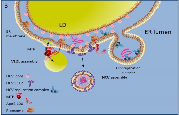
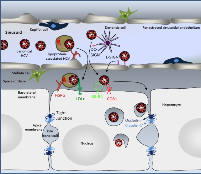
More than 170 million patients worldwide are chronically infected with hepatitis C virus (HCV). Prevalence rates range from 0.5% in Northern European countries to 28% in some areas of Egypt. HCV is hepatotropic, and in many countries chronic hepatitis C is a leading cause of liver disease including fibrosis, cirrhosis and hepatocellular carcinoma. HCV persists in 50-85% of infected patients, and once chronic infection is established, spontaneous clearance is rare. HCV is a member of the Flaviviridae family, in which it forms its own genus. Many lines of evidence suggest that the HCV life cycle displays many differences to that of other Flaviviridae family members. Some of these differences may be due to the close interaction of HCV with its host's lipid and particular triglyceride metabolism in the liver, which may explain why the virus can be found in association with lipoproteins in serum of infected patients. This review focuses on the molecular events underlying the HCV cell entry process and the respective roles of cellular co-factors that have been implied in these events. These include, among others, the lipoprotein receptors low density lipoprotein receptor and scavenger receptor BI, the tight junction factors occludin and claudin-1 as well as the tetraspanin CD81. We discuss the roles of these cellular factors in HCV cell entry and how association of HCV with lipoproteins may modulate the cell entry process.

Keywords: cell entry; hepatitis C virus.


Figures

Figure 1
Figure 1
VLDL and HCV assembly. A. VLDL assembly. Upon translation, nascent apolipoprotein 100 (apoB100) translocates through the endoplasmatic reticulum (ER) membrane. The protein is very unstable and subject to proteasomal degradation, unless it is rapidly lipidated. Lipidation of nascent apoB can occur by two different mechanisms. Either free MTP binds nascent apoB and subsequently extracts lipids from the membrane and transfers them into a hydrophobic pocket of the nascent apoB polypeptide. MTP molecules that are not physically associated with apoB can further assist this step and the pocket may serve as a nucleation site for lipid deposition. Several rounds of this process will result in extensive lipidation of apoB until the apolipoprotein/lipid complex is large enough to bud off from the rough ER membrane in the form of an immature VLDL. Alternatively, MTP associated with lipid vesicles or droplets may bind to apoB and provide a lipid core for the nascent apoB to encircle and wrap around it [11]. It is thought that the fusion of immature VLDL with lipidic vesicles derived from smooth ER regions (separation of rough and smooth ER membrane regions is indicated in the figure by ][) induces the maturation and secretion of VLDL from the hepatocytes into the blood. B. HCV assembly. HCV core protein localizes to the surface of lipid droplets and is able to interact with viral structural proteins assembled on the ER. Furthermore, intracellular membranes containing the HCV replication complex are enriched in MTP, apoB, and also apoE, and inhibition of the expression or activity of either of these factors blocks the release of infectious HCV. Thus, the release of infectious HCV may depend on virions being packaged as a VLDL-like particle.
Figure 1
Figure 1
VLDL and HCV assembly. A. VLDL assembly. Upon translation, nascent apolipoprotein 100 (apoB100) translocates through the endoplasmatic reticulum (ER) membrane. The protein is very unstable and subject to proteasomal degradation, unless it is rapidly lipidated. Lipidation of nascent apoB can occur by two different mechanisms. Either free MTP binds nascent apoB and subsequently extracts lipids from the membrane and transfers them into a hydrophobic pocket of the nascent apoB polypeptide. MTP molecules that are not physically associated with apoB can further assist this step and the pocket may serve as a nucleation site for lipid deposition. Several rounds of this process will result in extensive lipidation of apoB until the apolipoprotein/lipid complex is large enough to bud off from the rough ER membrane in the form of an immature VLDL. Alternatively, MTP associated with lipid vesicles or droplets may bind to apoB and provide a lipid core for the nascent apoB to encircle and wrap around it [11]. It is thought that the fusion of immature VLDL with lipidic vesicles derived from smooth ER regions (separation of rough and smooth ER membrane regions is indicated in the figure by ][) induces the maturation and secretion of VLDL from the hepatocytes into the blood. B. HCV assembly. HCV core protein localizes to the surface of lipid droplets and is able to interact with viral structural proteins assembled on the ER. Furthermore, intracellular membranes containing the HCV replication complex are enriched in MTP, apoB, and also apoE, and inhibition of the expression or activity of either of these factors blocks the release of infectious HCV. Thus, the release of infectious HCV may depend on virions being packaged as a VLDL-like particle.
Figure 2
Figure 2
HCV liver and cell entry. HCV circulates in the blood in canonical and lipoprotein-associated forms. To cross from the fenestrated sinusoidal liver endothelium into the space of Disse, HCV may diffuse through fenestrae or it may be captured and transcytosed by C-type lectins L-SIGN or DC-SIGN, expressed on the liver endothelium and dentritic cells, respectively. Alternatively, heparan sulfate proteoglycans (HSPG) on the surface of endothelial cells may favor the transfer of canonical and lipoprotein-associated virus into the liver. In the liver, canonical HCV hepatocytes entry requires the cellular entry factors scavenger receptor BI (SR-BI), the tetraspanin CD81 as well as Occludin and Claudin-1, tight junction factors that play a role in the physical separation of basolateral and apical membranes. HSPG and low density lipoprotein receptor (LDLr) have also been implied in HCV cell entry, but whether their roles apply particularly to lipoprotein-associated HCV remains unclear. HCV-virions are internalized into hepatocytes by clathrin-mediated endocytosis and overall the roles of the various HCV entry factors in cell attachment, internalization and fusion are not yet elucidated. Please refer to the text for further details.

References

    1. Alter MJ. Epidemiology of hepatitis C virus infection. World J Gastroenterol. 2007;13:2436–2441. - PMC - PubMed
    1. Moradpour D, Penin F, Rice CM. Replication of hepatitis C virus. Nat Rev Microbiol. 2007;5:453–463. - PubMed
    1. Lindenbach BD, Thiel HJ, Rice CM. Flaviviridae: The Viruses and Their Replication. In: Knipe DM, Howley PM, editors. Fields Virology. 5th ed. Vol. 1. Lippincott Williams & Wilkins; PA, USA: 2007. pp. 1101–1152.
    1. Negro F. Mechanisms and significance of liver steatosis in hepatitis C virus infection. World J Gastroenterol. 2006;12:6756–6765. - PMC - PubMed
    1. Miyanari Y, Atsuzawa K, Usuda N, Watashi K, Hishiki T, Zayas M, Bartenschlager R, Wakita T, Hijikata M, Shimotohno K. The lipid droplet is an important organelle for hepatitis C virus production. Nat Cell Biol. 2007;9:1089–1097. - PubMed

LinkOut - more resources