. 2010 Mar;2(3):710-730.
doi: 10.3390/v2030710. Epub 2010 Mar 9.

Complete genomic sequence of bacteriophage felix o1

Affiliations

Complete genomic sequence of bacteriophage felix o1

Jean M Whichard et al. Viruses. 2010 Mar.

Abstract

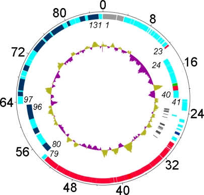
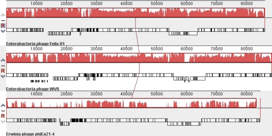
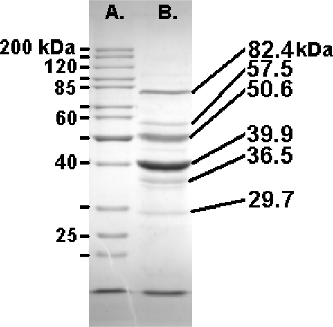
Bacteriophage O1 is a Myoviridae A1 group member used historically for identifying Salmonella. Sequencing revealed a single, linear, 86,155-base-pair genome with 39% average G+C content, 131 open reading frames, and 22 tRNAs. Closest protein homologs occur in Erwinia amylovora phage φEa21-4 and Escherichia coli phage wV8. Proteomic analysis indentified structural proteins: Gp23, Gp36 (major tail protein), Gp49, Gp53, Gp54, Gp55, Gp57, Gp58 (major capsid protein), Gp59, Gp63, Gp64, Gp67, Gp68, Gp69, Gp73, Gp74 and Gp77 (tail fiber). Based on phage-host codon differences, 7 tRNAs could affect translation rate during infection. Introns, holin-lysin cassettes, bacterial toxin homologs and host RNA polymerase-modifying genes were absent.

Keywords: DNA sequence; Felix O1; Myoviridae; Salmonella; bacteriophage; bioinformatics.


Figures

Figure 1
Figure 1
Genetic and physical map of phage Felix O1 prepared using DNAPlotter [20] with, from outer to inner rings, the size in kb; genes on the forward and reverse strands with some of the genes listed in italics (N.B. gene 1 = rIIA), and tRNA-encoding genes (black). The inner circles correspond to a GC plot (purple, below average GC-content; greenish-brown) and a GC skew analysis. Genes involved in nucleotide metabolism or DNA replication are indicated in dark blue, those involved in DNA packaging and morphogenesis in red; and, the lysis gene in dark green.
Figure 2
Figure 2
Mauve alignment of the genomes of phages Felix O1 (top), wV8 (middle) and φEa21-4 (bottom). The degree of DNA sequence similarity is indicated by the height of the red-coloured regions. Just above the phage names are box-like diagrams indicating the position of the genes.
Figure 3
Figure 3
SDS-PAGE (10%) analysis of the structural proteins of Felix O1 with the masses of the Fermentas PageRuler markers (left) and the most abundant proteins (right).

Similar articles

Cited by

References

    1. Ackermann HW. Tailed bacteriophages: the order. Caudovirales Adv Virus Res. 1998;51:135–201. - PMC - PubMed
    1. Ackermann HW. Salmonella Phages Observed in the Electron Microscope. In: Schatten H, Eisenstark A, editors. Salmonella Methods and Protocols. Humana Press; Totowa, NJ, USA: 2007. pp. 213–234.
    1. Kropinski AM, Sulakvelidze A, Konczy P, Poppe C. Salmonella Phages and Prophages - Genomics and Practical Aspects. In: Schatten H, Eisenstark A, editors. Salmonella Methods and Protocols. Humana Press; Totowa, NJ, USA: 2007. pp. 133–176. - PubMed
    1. Villafane R, Zayas M, Gilcrease EB, Kropinski AM, Casjens SR. Genomic analysis of bacteriophage ɛ34 of Salmonella enterica serovar Anatum (15+) BMC Microbiol. 2008;8:227. - PMC - PubMed
    1. Kwon HJ, Cho SH, Kim TE, Won YJ, Jeong J, Park SC, Kim JH, Yoo HS, Park YH, Kim SJ. Characterization of a T7-like lytic bacteriophage (phiSG-JL2) of Salmonella enterica serovar gallinarum biovar gallinarum. Appl Environ Microbiol. 2008;74:6970–6979. - PMC - PubMed

LinkOut - more resources