

Lipid membranes in poxvirus replication
- PMID: 21994664
- PMCID: PMC3185658
- DOI: 10.3390/v2040972
Lipid membranes in poxvirus replication
Abstract
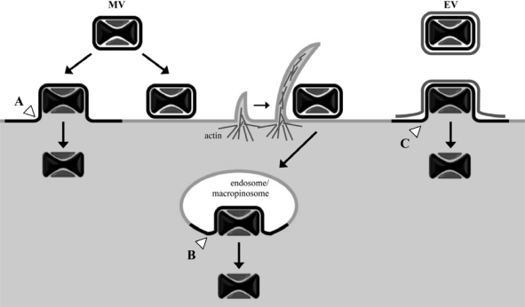
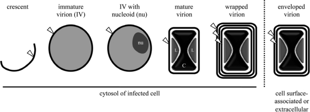
Poxviruses replicate in the cytoplasm, where they acquire multiple lipoprotein membranes. Although a proposal that the initial membrane arises de novo has not been substantiated, there is no accepted explanation for its formation from cellular membranes. A subsequent membrane-wrapping step involving modified trans-Golgi or endosomal cisternae results in a particle with three membranes. These wrapped virions traverse the cytoplasm on microtubules; the outermost membrane is lost during exocytosis, the middle one is lost just prior to cell entry, and the remaining membrane fuses with the cell to allow the virus core to enter the cytoplasm and initiate a new infection.
Keywords: endocytosis; exocytosis; phospholipids; transmembrane proteins; virus assembly; virus entry.
Figures
References
-
- Moss B. Poxviridae: the viruses and their replication. In: Knipe DM, editor. Fields Virology. 5th ed. Vol. 2 Lippincott Williams & Wilkins; Philadelphia, PA, USA: 2007.
-
- Fenner F, Henderson DA, Arita I, Jezek Z, Ladnyi ID, editors. Smallpox and Its Eradication. World Health Organization; Geneva, Switzerland: 1988. Development of the global smallpox eradication programme, 1958–1966; pp. 365–419.
-
- Condit RC, Moussatche N, Traktman P. In a nutshell: structure and assembly of the vaccinia virion. Adv Virus Res. 2006;66:31–124. - PubMed
LinkOut - more resources
Full Text Sources
Other Literature Sources
