. 2010 Apr;2(4):1011-1049.
doi: 10.3390/v2041011. Epub 2010 Apr 15.

Glycosphingolipids as receptors for non-enveloped viruses

Affiliations

Glycosphingolipids as receptors for non-enveloped viruses

Stefan Taube et al. Viruses. 2010 Apr.

Abstract

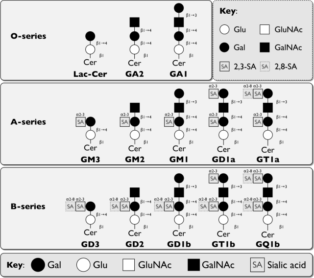
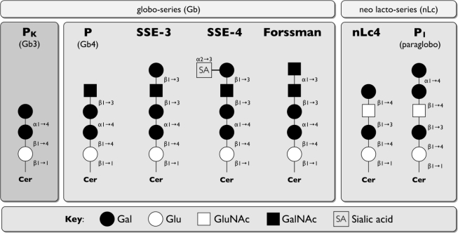
Glycosphingolipids are ubiquitous molecules composed of a lipid and a carbohydrate moiety. Their main functions are as antigen/toxin receptors, in cell adhesion/recognition processes, or initiation/modulation of signal transduction pathways. Microbes take advantage of the different carbohydrate structures displayed on a specific cell surface for attachment during infection. For some viruses, such as the polyomaviruses, binding to gangliosides determines the internalization pathway into cells. For others, the interaction between microbe and carbohydrate can be a critical determinant for host susceptibility. In this review, we summarize the role of glycosphingolipids as receptors for members of the non-enveloped calici-, rota-, polyoma- and parvovirus families.

Keywords: calicivirus; glycosphingolipid; non-enveloped virus; parvovirus; polyomavirus; receptor; rotavirus.


Figures

Figure 1.
Figure 1.
Schematic representation of discussed major core structures of vertebrate glycosphingolipids (GSLs) (based on IUPAC-IUB Joint Commission on Biochemical Nomenclature [6]). Abbreviations: Glu: glucose, Gal: galactose, GluNAc: N-acetylglucosamine, GalNAc: N-acetylgalactosamine, Cer: ceramide, R: residue
Figure 2.
Figure 2.
The biosynthetic pathway of type 1 (Lc) and type 2 (nLc) HBGAs. The pathway starts with a disaccharide precursor of type1 or type 2 carbohydrate chains. Type 1 disaccharide precursors are β1-3, and type 2 are β1-4 linked. FUT1 specifically generates type 2 H antigens, while FUT2 preferentially produces the type 1 H antigens, although it also has activity on the type 2 chain [109]. Further modification of the H type1/2 by A or B enzymes synthesizes the respective tetrasaccharides, adding the A and B antigens. FUT3 generates the trisaccharides Lewis A (LeA) or Lewis X (LeX) in non-secretors, as well as LeB and LeY in secretors, by attaching α1-4 fucose on type 1 chains or α1-3 fucose on type 2 chains. Abbreviations: Glu: glucose, Gal: galactose, GluNAc: N-acetylglucosamine, GalNAc: N-acetylgalactosamine, Cer: ceramide, R: residue
Figure 3.
Figure 3.
Comparison of some discussed carbohydrate structures used during attachment of human and mouse NoV. Schematic representation of the HuNoV attachment receptors (H type 1, H type 3 A, Sialyl Lewis X) and MNV-1 ganglioside attachment receptors (GD1a and GT1b). Abbreviations: Glu: glucose, Gal: galactose, GluNAc: N-acetylglucosamine, GalNAc: N-acetylgalactosamine, Cer: ceramide, R: residue
Figure 4.
Figure 4.
Comparison of discussed carbohydrate structures used during attachment of bovine NoV. Schematic representation of the BoNoV attachment receptor (αGal and αGal-LeX) in comparison to the human HBGA receptor H type 2 B-antigen. Abbreviations: Glu: glucose, Gal: galactose, GluNAc: N-acetylglucosamine, GalNAc: N-acetylgalactosamine, Cer: ceramide, R: residue
Figure 5.
Figure 5.
Schematic overview of O-, A- and B-series gangliosides discussed in text. Abbreviations: Glu: glucose, Gal: galactose, GluNAc: N-acetylglucosamine, GalNAc: N-acetylgalactosamine, Cer: ceramide, R: residue
Figure 6.
Figure 6.
Comparison of discussed carbohydrate structures used during attachment of parvovirus B19. Schematic representation of nLC and Gb attachment receptors in comparison to the non-binding globoside Gb3. Abbreviations: Glu: glucose, Gal: galactose, GluNAc: N-acetylglucosamine, GalNAc: N-acetylgalactosamine, Cer: ceramide, R: residue

Similar articles

Cited by

References

    1. Helenius A. Virus entry and uncoating. In: Knipe DM, Howley PM, editors. Fields Virology. 5 ed. Vol. 1. Wolters Kluwer; Philadelphia: 2007. pp. 99–118.
    1. Olofsson S, Bergstrom T. Glycoconjugate glycans as viral receptors. Ann Med. 2005;37:154–172. - PubMed
    1. Iwamori M. A new turning point in glycosphingolipid research. Hum Cell. 2005;18:117–133. - PubMed
    1. Prinetti A, Loberto N, Chigorno V, Sonnino S. Glycosphingolipid behaviour in complex membranes. Biochim Biophys Acta. 2009;1788:184–193. - PubMed
    1. Hakomori S. Structure, organization, and function of glycosphingolipids in membrane. Curr Opin Hematol. 2003;10:16–24. - PubMed

LinkOut - more resources