

The HTLV-1 Virological Synapse
- PMID: 21994688
- PMCID: PMC3185711
- DOI: 10.3390/v2071427
The HTLV-1 Virological Synapse
Abstract
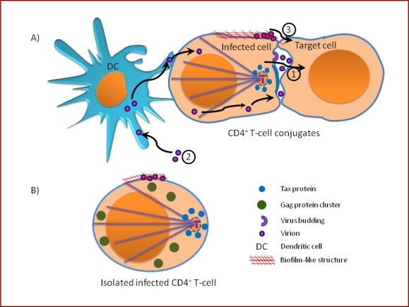
Human T-lymphotropic virus-1 (HTLV-1) spreads efficiently between T-cells via a tight and highly organized cell-cell contact known as the virological synapse. It is now thought that many retroviruses and other viruses spread via a virological synapse, which may be defined as a virus-induced, specialized area of cell-to-cell contact that promotes the directed transmission of the virus between cells. We summarize here the mechanisms leading to the formation of the HTLV-1 virological synapse and the role played by HTLV-1 Tax protein. We propose a model of HTLV-1 transmission between T-cells based on the three-dimensional ultrastructure of the virological synapse. Finally, in the light of recent advances, we discuss the possible routes of HTLV-1 spread across the virological synapse.
Keywords: HTLV-1 virological synapse; cytoskeleton; polarization; viral proteins.
Figures
Similar articles
-
Human T-lymphotropic virus, type 1, tax protein triggers microtubule reorientation in the virological synapse.J Biol Chem. 2005 Aug 19;280(33):29653-60. doi: 10.1074/jbc.M502639200. Epub 2005 Jun 23. J Biol Chem. 2005. PMID: 15975923
-
HTLV-1-Tax and ICAM-1 act on T-cell signal pathways to polarize the microtubule-organizing center at the virological synapse.Blood. 2009 Jul 30;114(5):1016-25. doi: 10.1182/blood-2008-03-136770. Epub 2009 Jun 3. Blood. 2009. PMID: 19494354
-
Human T-lymphotropic virus-1 visualized at the virological synapse by electron tomography.PLoS One. 2008 May 28;3(5):e2251. doi: 10.1371/journal.pone.0002251. PLoS One. 2008. PMID: 18509526 Free PMC article.
-
[Intercellular transmission of HTLV-1: not all mechanisms have been revealed].Med Sci (Paris). 2015 Jun-Jul;31(6-7):629-37. doi: 10.1051/medsci/20153106016. Epub 2015 Jul 7. Med Sci (Paris). 2015. PMID: 26152167 Review. French.
-
Molecular Mechanisms of HTLV-1 Cell-to-Cell Transmission.Viruses. 2016 Mar 9;8(3):74. doi: 10.3390/v8030074. Viruses. 2016. PMID: 27005656 Free PMC article. Review.
Cited by
-
Determination of molecular epidemiologic pattern of human T-lymphotropic virus type 1 (HTLV-1) in Alborz province, Iran.Virus Genes. 2024 Apr;60(2):117-125. doi: 10.1007/s11262-024-02051-0. Epub 2024 Jan 25. Virus Genes. 2024. PMID: 38273115
-
Molecular determinants of the ratio of inert to infectious virus particles.Prog Mol Biol Transl Sci. 2015;129:285-326. doi: 10.1016/bs.pmbts.2014.10.012. Epub 2014 Dec 1. Prog Mol Biol Transl Sci. 2015. PMID: 25595808 Free PMC article. Review.
-
The Assembly of HTLV-1-How Does It Differ from HIV-1?Viruses. 2024 Sep 27;16(10):1528. doi: 10.3390/v16101528. Viruses. 2024. PMID: 39459862 Free PMC article. Review.
-
Cell surface markers in HTLV-1 pathogenesis.Viruses. 2011 Aug;3(8):1439-59. doi: 10.3390/v3081439. Epub 2011 Aug 16. Viruses. 2011. PMID: 21994790 Free PMC article. Review.
-
Cell-to-cell transmission of retroviruses: Innate immunity and interferon-induced restriction factors.Virology. 2011 Mar 15;411(2):251-9. doi: 10.1016/j.virol.2010.12.031. Epub 2011 Jan 17. Virology. 2011. PMID: 21247613 Free PMC article. Review.
References
-
- Uchiyama T, Yodoi J, Sagawa K, Takatsuki K, Uchino H. Adult T-cell leukemia: clinical and hematologic features of 16 cases. Blood. 1977;50:481–492. - PubMed
-
- Gessain A, Barin F, Vernant JC, Gout O, Maurs L, Calender A, de The G. Antibodies to human T-lymphotropic virus type-I in patients with tropical spastic paraparesis. Lancet. 1985;2:407–410. - PubMed
-
- Osame M, Izumo S, Igata A, Matsumoto M, Matsumoto T, Sonoda S, Tara M, Shibata Y. Blood transfusion and HTLV-I associated myelopathy. Lancet. 1986;2:104–105. - PubMed
-
- Wattel E, Cavrois M, Gessain A, Wain-Hobson S. Clonal expansion of infected cells: a way of life for HTLV-I. J Acquir Immune Defic Syndr Hum Retrovirol. 1996;13:S92–99. - PubMed
-
- Richardson JH, Hollsberg P, Windhagen A, Child LA, Hafler DA, Lever AM. Variable immortalizing potential and frequent virus latency in blood-derived T-cell clones infected with human T-cell leukemia virus type I. Blood. 1997;89:3303–3314. - PubMed
LinkOut - more resources
Full Text Sources
