. 2010 Sep;2(9):1933-1967.
doi: 10.3390/v2091933. Epub 2010 Sep 15.

Orthopoxvirus genome evolution: the role of gene loss

Affiliations

Orthopoxvirus genome evolution: the role of gene loss

Robert Curtis Hendrickson et al. Viruses. 2010 Sep.

Abstract

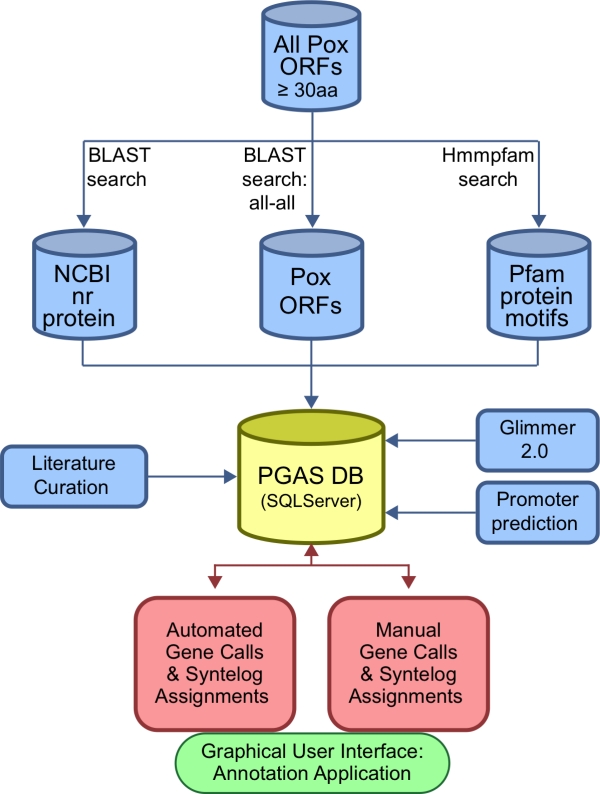
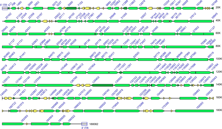
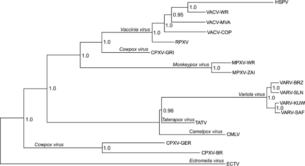
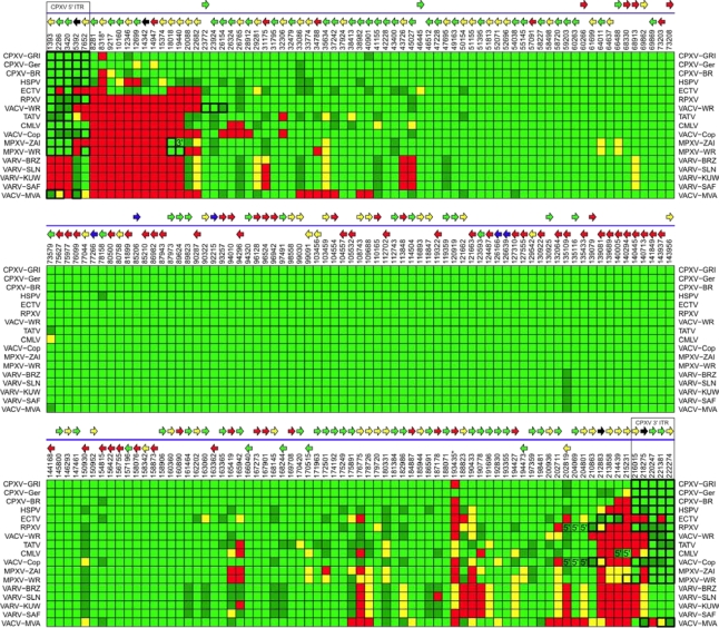
Poxviruses are highly successful pathogens, known to infect a variety of hosts. The family Poxviridae includes Variola virus, the causative agent of smallpox, which has been eradicated as a public health threat but could potentially reemerge as a bioterrorist threat. The risk scenario includes other animal poxviruses and genetically engineered manipulations of poxviruses. Studies of orthologous gene sets have established the evolutionary relationships of members within the Poxviridae family. It is not clear, however, how variations between family members arose in the past, an important issue in understanding how these viruses may vary and possibly produce future threats. Using a newly developed poxvirus-specific tool, we predicted accurate gene sets for viruses with completely sequenced genomes in the genus Orthopoxvirus. Employing sensitive sequence comparison techniques together with comparison of syntenic gene maps, we established the relationships between all viral gene sets. These techniques allowed us to unambiguously identify the gene loss/gain events that have occurred over the course of orthopoxvirus evolution. It is clear that for all existing Orthopoxvirus species, no individual species has acquired protein-coding genes unique to that species. All existing species contain genes that are all present in members of the species Cowpox virus and that cowpox virus strains contain every gene present in any other orthopoxvirus strain. These results support a theory of reductive evolution in which the reduction in size of the core gene set of a putative ancestral virus played a critical role in speciation and confining any newly emerging virus species to a particular environmental (host or tissue) niche.

Keywords: bioinformatics; evolution; orthopoxviruses; poxviruses; variola virus.


Figures

Figure 1.
Figure 1.
Gene sequence phylogeny of the family Poxviridae. Phylogenetic prediction based on an amino acid alignment of 20 conserved genes from representative virus isolates. Each terminal node is labeled with the genus name; and the type species for each genus is provided in parentheses. Unclassified viruses have not yet been assigned to a taxon. The Orthopoxvirus genus, analyzed in this manuscript, is highlighted.
Figure 2.
Figure 2.
The Poxvirus Genome Annotation System (PGAS) design. The PGAS pipeline (blue) automatically runs the underlying analyses in parallel on a local high-performance computing cluster for each new genome. Results from those analyses are then loaded into the PGAS database (yellow). The process of making gene calls (red) is directed from a desktop java GUI application (green).
Figure 3.
Figure 3.
PGAS Screen Shots (a) Single genome display for HSPV and a dual-genome display of the same HSPV region and its homolog in CPXV-GRI. ORFs from all 6 translational reading frames are shown as horizontal lines, with putative, and as available, verified transcriptional promoters indicated by colored vertical lines. ORFs showing similarity by BLAST analysis are connected by red lines. ORFs that have been annotated into the same orthologous gene family are connected by thicker lines. (b) Results of the BLAST search between two ORFs (left panel). The results of a Needleman-Wunsch (global) alignment is displayed in the right panel. (c) Multi-genome homolog comparison of a representative ORF showing the predicted start of the coding region with potential ATG translation start sites highlighted. The 5′ untranslated region with predicted promoter sequences highlighted is also displayed. Early termination sequences from the upstream gene are indicated in red. Late promoters are colored pink. (d) Homologs across multiple genomes can also be displayed using a BLAST/Pfam viewer. Examples of fragmented (left panel) and truncated (right panel) genes are shown.
Figure 3.
Figure 3.
PGAS Screen Shots (a) Single genome display for HSPV and a dual-genome display of the same HSPV region and its homolog in CPXV-GRI. ORFs from all 6 translational reading frames are shown as horizontal lines, with putative, and as available, verified transcriptional promoters indicated by colored vertical lines. ORFs showing similarity by BLAST analysis are connected by red lines. ORFs that have been annotated into the same orthologous gene family are connected by thicker lines. (b) Results of the BLAST search between two ORFs (left panel). The results of a Needleman-Wunsch (global) alignment is displayed in the right panel. (c) Multi-genome homolog comparison of a representative ORF showing the predicted start of the coding region with potential ATG translation start sites highlighted. The 5′ untranslated region with predicted promoter sequences highlighted is also displayed. Early termination sequences from the upstream gene are indicated in red. Late promoters are colored pink. (d) Homologs across multiple genomes can also be displayed using a BLAST/Pfam viewer. Examples of fragmented (left panel) and truncated (right panel) genes are shown.
Figure 4.
Figure 4.
Fragmentation of orthopoxvirus genes (a) Fragmentation pattern of the guanylate kinase gene. (b) Fragmentation pattern of the A-type inclusion protein. Virus abbreviations are defined in Table 1.
Figure 5.
Figure 5.
Variola virus strain Brazil 1966 Genome Map. Each arrow indicates the presence of an ORF within the VARV-BRZ genome. The arrow also designates the direction of transcription. Intact genes are colored using light green arrows; truncated genes by dark green arrows; and fragmented genes by yellow arrows. The numerical designations indicate the position of the last base of each ORF. The position of the ITRs at both ends of the genome is also indicated.
Figure 6.
Figure 6.
Gene sequence phylogeny of the genus Orthopoxvirus. Codon-aligned gene sequences of 141 genes from each indicated orthopoxvirus were used for phylogenetic prediction using Bayesian inference. Species names are indicated along the branch distinguishing each species clade, and strain names are provided at each terminal node. The numbers at each node provide the clade credibility values for each node—a measure of the confidence of the branching pattern for the indicated clade.
Figure 7.
Figure 7.
Comparison of orthopoxvirus gene lengths. The length of every annotated orthopoxvirus gene from the genomes listed in Table 1 was compared to the length of the corresponding cowpox virus ortholog. The length of each gene as a percentage of the length of the longest cowpox virus strain gene was plotted with respect to its genomic position in CPXV-GRI.
Figure 8.
Figure 8.
Comparative orthopoxvirus gene conservation map.
Figure 9.
Figure 9.
Gene loss summary. The number of intact (light green), truncated (dark green), fragmented (yellow), and missing (red) gene families is plotted for each virus strain.
Figure 10.
Figure 10.
Gene content phylogeny of the genus Orthopoxvirus. A Bayesian phylogenetic tree inferred on the basis of similarities in gene content between virus strains. Strain names are provided at each terminal node. The numbers at each branch point provide the clade credibility values for each node—a measure of the confidence of the branching pattern for the indicated clade.

References

    1. Odom MR, Hendrickson RC, Lefkowitz EJ. Poxvirus protein evolution: Family wide assessment of possible horizontal gene transfer events. Virus Res. 2009;144:233–249. - PMC - PubMed
    1. Fauquet CM, Mayo MA, Maniloff J, Desselberger U, Ball LA. Virus Taxonomy: VIIIth Report of the International Committee on Taxonomy of Viruses. Academic Press (Elsevier); London, UK: 2005.
    1. Mercer AA, Schmidt A, Weber OF. Poxviruses. Birkhäuser Verlag; Basel, Switzerland: 2007.
    1. Barrett JW, McFadden G. Origin and evolution of poxviruses. In: Domingo E, Parrish CR, Holland JJ, editors. Origin and Evolution of Viruses. 2nd ed. Academic Press (Elsevier); London, UK: 2008. pp. 431–446.
    1. DeFilippis VR, Villarreal LP. Virus evolution. In: Knipe DM, Howley PM, editors. Fields Virology. 4th ed. Lippincott Williams & Wilkins; Philadelphia, PA, USA: 2001. pp. 353–370.

LinkOut - more resources