

Review
doi: 10.3390/v3030172.
Epub 2011 Feb 25.
Bacteriophage assembly
Affiliations
- PMID: 21994726
- PMCID: PMC3185693
- DOI: 10.3390/v3030172
Item in Clipboard
Review
Bacteriophage assembly
Viruses.
2011 Mar.
Abstract
Bacteriophages have been a model system to study assembly processes for over half a century. Formation of infectious phage particles involves specific protein-protein and protein-nucleic acid interactions, as well as large conformational changes of assembly precursors. The sequence and molecular mechanisms of phage assembly have been elucidated by a variety of methods. Differences and similarities of assembly processes in several different groups of bacteriophages are discussed in this review. The general principles of phage assembly are applicable to many macromolecular complexes.
Keywords: assembly; bacteriophage; genome packaging; macromolecular complex; maturation.
Figures
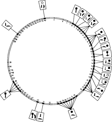
A simplified bacteriophage T4 genome map showing the effect of mutations in morphological genes on virion assembly. Boxed symbols attached to a particular gene show the phenotype, arising from the defects in that gene. Reprinted from Wood et al. [1]; with permission. Copyright for this figure is owned by the Federation of American Societies for Experimental Biology. This image may not be reproduced in any way without explicit permission from the copyright owner.
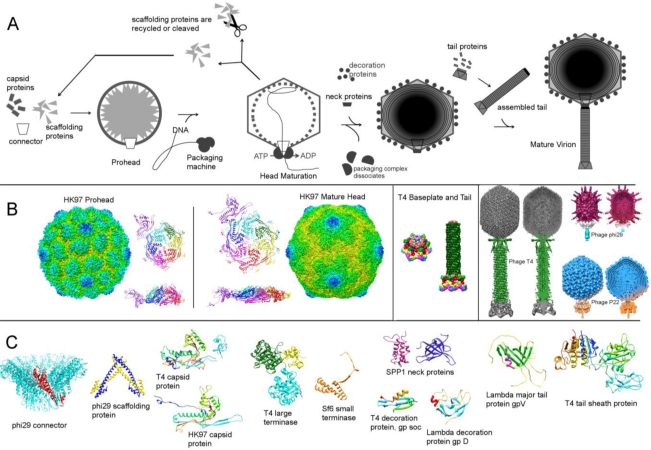
Assembly of dsDNA tailed phages. (A) Schematic of the assembly steps. (B) Examples of assembly intermediates and mature virions. The left panel shows HK97 prohead and mature head, displaying the whole structure, as well as side and top views of the icosahedral asymmetric units. The middle panel shows the T4 baseplate and tail. The right panel shows T4, phi29 and P22 mature virions, as a surface rendered view and a slab view, displaying the DNA structure. (C) Structural proteins involved in the assembly of several phages. The figure was created using the program CHIMERA [180]. The relevant structures were downloaded from the PDB and EMDB databases.
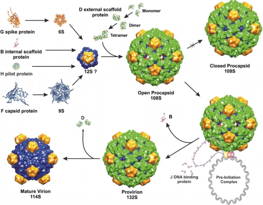
Microviridae assembly pathway (reprinted from Bernal et al. [189] with permission from Elsevier).
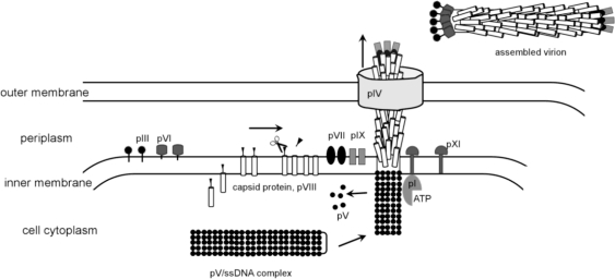
Model for the assembly and extrusion of filamentous phages. The figure was created based on a figure by Russel and Model [196].
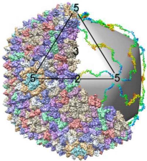
Structure of PRD1 virion. A portion of the capsid protein was removed to show the mesh underneath made by 60 copies of tape measure protein p30. The figure was created using the program CHIMERA [180]. The relevant PDB entry 1W8X was downloaded from the PDB databank.
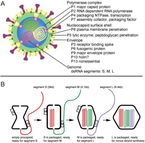
(A) A diagram showing the phi6 virion architecture (reprinted from [228] with permission from Elsevier). (B) Genome packaging in dsRNA phages (reprinted from Figure 6 in Mindich et al. [16] with permission from Elsevier).
References
-
- Wood WB, Edgar RS, King J, Lielausis I, Henninger M. Bacteriophage assembly. Fed. Proc. 1968;27:1160–1166. - PubMed
-
- Casjens S, King J. Virus assembly. Annu. Rev. Biochem. 1975;44:555–611. - PubMed
-
- Bazinet C, King J. The DNA translocating vertex of dsDNA bacteriophage. Annu. Rev. Microbiol. 1985;39:109–129. - PubMed
-
- Black LW. DNA packaging in dsDNA bacteriophages. Annu. Rev. Microbiol. 1989;43:267–292. - PubMed
Publication types
MeSH terms
Substances
Grants and funding
LinkOut - more resources
Full Text Sources
