. 2011 Jul;3(7):1041-1058.
doi: 10.3390/v3071041. Epub 2011 Jul 5.

C7a, a biphosphinic cyclopalladated compound, efficiently controls the development of a patient-derived xenograft model of adult T cell leukemia/lymphoma

Affiliations

C7a, a biphosphinic cyclopalladated compound, efficiently controls the development of a patient-derived xenograft model of adult T cell leukemia/lymphoma

Ana B Guimaraes-Correa et al. Viruses. 2011 Jul.

Abstract

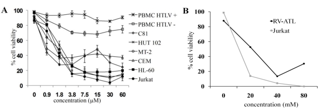
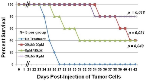
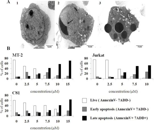
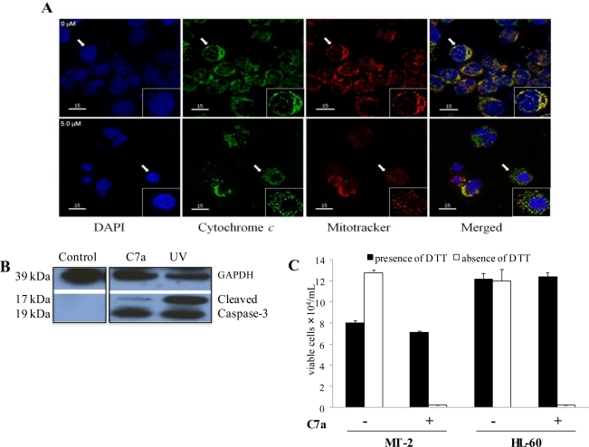
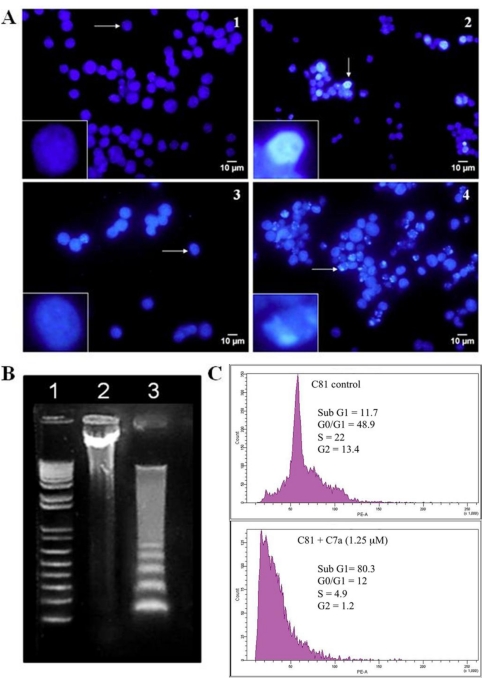
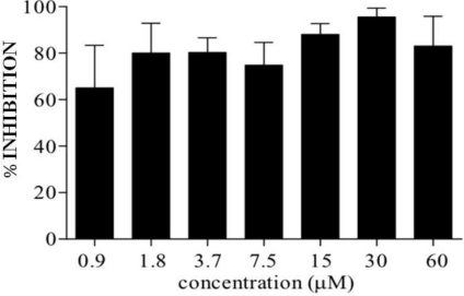
Adult T-cell leukemia/lymphoma (ATLL) is a highly aggressive disease that occurs in individuals infected with the human T lymphotropic virus type 1 (HTLV-1). Patients with aggressive ATLL have a poor prognosis because the leukemic cells are resistant to conventional chemotherapy. We have investigated the therapeutic efficacy of a biphosphinic cyclopalladated complex {Pd(2) [S(-)C(2), N-dmpa](2) (μ-dppe)Cl(2)}, termed C7a, in a patient-derived xenograft model of ATLL, and investigated the mechanism of C7a action in HTLV-1-positive and negative transformed T cell lines in vitro. In vivo survival studies in immunocompromised mice inoculated with human RV-ATL cells and intraperitoneally treated with C7a led to significantly increased survival of the treated mice. We investigated the mechanism of C7a activity in vitro and found that it induced mitochondrial release of cytochrome c, caspase activation, nuclear condensation and DNA degradation. These results suggest that C7a triggers apoptotic cell death in both HTLV-1 infected and uninfected human transformed T-cell lines. Significantly, C7a was not cytotoxic to peripheral blood mononuclear cells (PBMC) from healthy donors and HTLV-1-infected individuals. C7a inhibited more than 60% of the ex vivo spontaneous proliferation of PBMC from HTLV-1-infected individuals. These results support a potential therapeutic role for C7a in both ATLL and HTLV-1-negative T-cell lymphomas.

Keywords: ATLL; HTLV-1; apoptosis; chemotherapy; cyclopalladated compound; xenograft model.


Figures

Figure 1
Figure 1
Cyclopalladated C7a is cytotoxic to human leukemia cell lines, but has little effect on human T lymphotropic virus type 1 (HTLV-1)-infected or uninfected peripheral blood mononuclear cells (PBMC). (A) 5 × 10⁵ PBMC (previously stimulated with PHA and IL-2) from three donors (HTLV-1 uninfected), 105 cells from human T cell leukemia lines, and 105 PBMC from four different HTLV-1-infected individuals were seeded in 96-well plates in triplicate. Cells were incubated with concentrations of C7a as indicated for 48 h, and cytotoxicity was determined by Trypan blue exclusion in an automated cell counter. The bars represent the means and SD of three different experiments; (B) RV-ATL cells were expanded in a SCID mouse for three weeks. Freshly harvested cells were seeded at 105 cells per well in a 24-well plate. The cells were incubated with concentrations of C7a as indicated for 48 h. Uninfected human Jurkat T cells were used as a control and incubated in the same conditions. Cytotoxicity was determined by Trypan blue exclusion and manual counting. A representative experiment is shown. The experiment was repeated twice, using two independent frozen tissue culture stocks of RV-ATL cells and without refreshing the media or drug, with comparable results. In (A) and (B), the viability of treated cells was expressed as percent of control untreated cells.
Figure 2
Figure 2
C7a significantly increases the survival of RV-ATL tumor bearing mice. Adult female NSG mice were intraperitoneally (IP) injected with 107 fresh, in vivo expanded RV-ATL cells on Day 0. Mice were treated with either C7a (20 μM diluted in PBS) (red line and purple line) or PBS alone (blue line and green line) by IP injection every other day starting on Day 4. Starting on day 22, half the mice from each group were treated with 80 μM C7a (green line and purple line). N = 5 per group for four treatment groups. All mice were treated by IP injections through day 40, and survival was monitored daily. p values (Kaplan Meier test) are shown for each group, compared to no treatment group.
Figure 3
Figure 3
C7a induces morphological and phenotypic alterations compatible with apoptosis in human leukemia cell lines. (A) Transmission electron microscopy. (1) HL-60 cells cultivated in complete RPMI medium for 6 h; (2 and 3) HL-60 cells cultivated in presence of 2 μM C7a for 7 h. Black arrows: nuclear condensation; White arrows: vacuoles. This assay was repeated twice, and representative images are shown; (B) Membrane phosphatidylserine expression. HTLV-1 infected MT-2 and C81, and uninfected Jurkat cell lines (1 × 10⁶) were incubated in the presence of C7a at the indicated concentrations for 3 h at 37 °C. After staining with PE-Annexin V and 7-AAD, cells were analyzed by flow cytometry, as described in Material and Methods. A representative of two independent experiments is shown.
Figure 4
Figure 4
Mitochondria are involved in C7a induced cell death. (A) Cytochrome c release. HTLV-1 infected MT-2 cells were incubated without (0 μM) or with 5 μM C7a for 3 h. The cells were fixed and incubated with anti-cytochrome c antibody (green). Mitochondria were detected by MitoTracker staining (red), and nuclei were visualized with DAPI (blue). Magnification (×40). Inserts: Single cell indicated by the arrow, magnification (×60). Rare cells treated with C7a that show colocalization of cytochrome c and mitochondria likely reflect the short 3 h exposure to C7a. The assay was repeated twice, and representative images are shown; (B) Caspase Activation. Cell lysates from MT-2 cells before treatment (Control), after treatment with 5 μM C7a for 3 h, or after exposure to 50 J of UV light (UV), as a positive control, were analyzed by Western blotting for cleaved caspase-3 and GAPDH as a loading control. This assay was repeated twice, and representative images are shown; (C) DTT inhibits C7a effect on leukemic human T cells. MT-2 (HTLV-1 infected) and uninfected HL-60 cells (105) were pre-incubated or not with 1 mM of DTT for 1 h. C7a was added to the culture (15 μM to MT-2, 2 μM to HL-60 cells) and incubated for 6 h. Viable cells were counted in presence of Trypan Blue in a hemocytometer. The bars represent the means and SD of three different experiments.
Figure 5
Figure 5
C7a induces nuclear alterations characteristic of apoptosis. (A) Nuclear condensation. Uninfected HL-60 cells were untreated (1) or treated with 5 μM C7a (2) for 6 h. Alternatively, HL-60 cells were untreated (3) or treated with 2.5 μM C7a (4) for 24 h. After incubation with C7a, cell nuclei were stained with Hoechst 33342. Inserts: Single cell indicated by the arrow, ×25 magnification. The assay was repeated three times, and representative images are shown; (B) DNA fragmentation. HL-60 cells (3 × 10⁶) were treated (lane 3) with 5 μM of C7a or not (lane 2) for 18 h. Lane 1: MW standard. The assay was repeated three times, and a representative gel is shown; (C) Cell cycle analysis. HTLV-1 infected C81 cells were cultured with 1.25 μM C7a (bottom panel) or not (top panel) for 48 h. Cells were stained with propidium iodide (50 μg/mL) and cell cycle analysis was performed by flow cytometry. Percentage of cells in each phase of the cycle is shown.
Figure 6
Figure 6
In vitro inhibition of spontaneous proliferation of PBMC from HTLV-1-infected patients by C7a. Isolated PBMC from HTLV-1-infected individuals were cultured in the presence or absence of C7a compound for 24 h. Bars represent average inhibition and standard deviation from four patients, compared to cells from the same patient cultivated in the absence of C7a.

References

    1. Nasr R, El Hajj H, Kfour Y, de Thé H, Hermine O, Bazarbachi A. Controversies in targeted therapy of adult T cell leukemia/lymphoma: ON target or off target effects? Viruses. 2011;3 in press. - PMC - PubMed
    1. Feuer G, Zack JA, Harrington WJ, Jr, Valderama R, Rosenblatt JD, Wachsman W, Baird SM, Chen IS. Establishment of HTLV- I T-cell lymphomas in severe combined immunodeficient mice. Blood. 1993;82:722–731. - PubMed
    1. Feuer G, Stewart SA, Baird SM, Lee F, Feuer G, Chen IS. Potential role of natural killer cells in controlling tumorigenesis by HTLVs. J. Virol. 1995;69:1328–1333. - PMC - PubMed
    1. Shultz LD, Lyons BL, Burzenski LM, Gott B, Chen X, Chaleff S, Kotb M, Gillies SD, King M, Mangada J, et al. Human lymphoid and myeloid cell development in NOD/LtSz-scid IL2R gamma null mice engrafted with mobilized human hemopoietic stem cells. J. Immunol. 2005;174:6477–6489. - PubMed
    1. Shultz LD, Ishikawa F, Greiner DL. Humanized mice in translational biomedical research. Nat. Rev. Immunol. 2007;7:118–130. - PubMed

Publication types

MeSH terms