Review
. 2011 Jul;3(7):1091-111.
doi: 10.3390/v3071091. Epub 2011 Jul 8.

The natural killer cell cytotoxic function is modulated by HIV-1 accessory proteins

Affiliations
Review

The natural killer cell cytotoxic function is modulated by HIV-1 accessory proteins

Bharatwaj Sowrirajan et al. Viruses. 2011 Jul.

Abstract

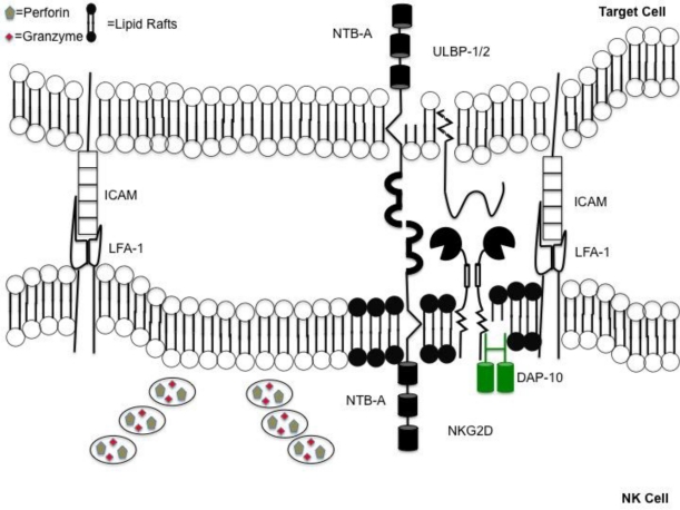
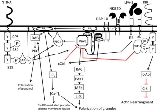
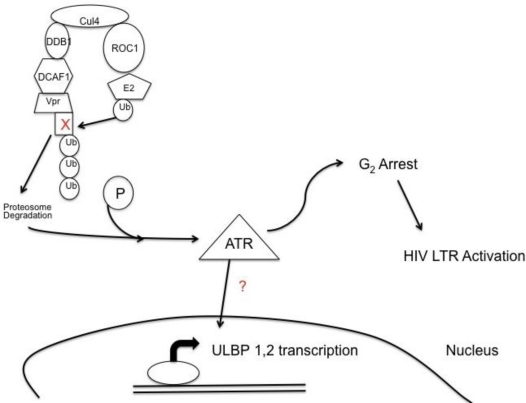
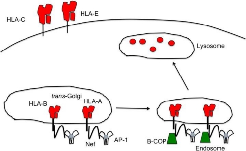
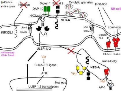
Natural killer (NK) cells' major role in the control of viruses is to eliminate established infected cells. The capacity of NK cells to kill virus-infected cells is dependent on the interactions between ligands on the infected cell and receptors on the NK cell surface. Because of the importance of ligand-receptor interactions in modulating the NK cell cytotoxic response, HIV has developed strategies to regulate various NK cell ligands making the infected cell surprisingly refractory to NK cell lysis. This is perplexing because the HIV-1 accessory protein Vpr induces expression of ligands for the NK cell activating receptor, NKG2D. In addition, the accessory protein Nef removes the inhibitory ligands HLA-A and -B. The reason for the ineffective killing by NK cells despite the strong potential to eliminate infected cells is due to HIV-1 Vpu's ability to down modulate the co-activation ligand, NTB-A, from the cell surface. Down modulation of NTB-A prevents efficient NK cell degranulation. This review will focus on the mechanisms through which the HIV-1 accessory proteins modulate their respective ligands, and its implication for NK cell killing of HIV-infected cells.

Keywords: HIV-1; NK cells; NKG2D; NTB-A; Nef; PLC-γ; Vpr; Vpu.


Figures

Figure 1
Figure 1
Coordinated events occur when forming an immunological synapse. Directed release of cytotoxic granules requires coordinated series of events. Synapse formation is accomplished following engagement of lymphocyte function-associated antigen (LFA)-1 on natural killer (NK) cells with intercellular adhesion molecules (ICAMs) on the target. Immunological synapse contains a “lipid raft” domain (shown in black) containing activating receptors. This is adjacent to a section of the plasma membrane allowing for the unobstructed release of perforin and granzymes, into the space between the NK cell and target.
Figure 2
Figure 2
Two activating signals are required to initiate NK cell degranulation. Signals from activating (NKG2D) and co-activating receptor (NTB-A) converge to activate phospholipase-Cγ (PLCγ). Vav1, a guanine exchange factor, is able to phosphorylate PLCγ when triggered by activating receptors. However, engagement of activating receptors also phosphorylates c-Cbl, an ubiquitin ligase that negatively regulates Vav1. To overcome this inhibition, a co-activating receptor must be engaged to phosphorylate sufficient PLCγ to trigger degranulation. Degranulation is mediated by the cleavage of phosphatidylinositol into diacylglycerol (DAG) and inositol 1,4,5 triphosphate (IP3). DAG recruits key proteins to the plasma membrane to initiate fusion of the granule and plasma membrane. IP3, once cleaved, binds its receptor, IP3 receptor, on the membrane of the endoplasmic reticulum to open calcium channels. Ca2+ influx allows for the exocytosis of the cytolytic granules. Inhibitory receptors such as the KIRs can recruit phosphatases that lead to the dephosphorylation of Vav1. However, the KIRs also have the ability to activate tyrosine kinases like c-Abl leading to the inhibition of actin rearrangement.
Figure 3
Figure 3
HIV-1 Vpr leads to expression of NKG2D ligands, unique long 16-binding protein (ULBP)-1 and -2. Viral protein R, Vpr, bridges DCAF1, a substrate recognition factor for an E3 ubiquitin ligase system to an unknown cell cycle protein causing for the cell cycle protein to become ubiquitinated and destroyed. The degradation of the cell cycle protein induces phosphorylation of the DNA damage sensor ataxia telangiectasia-mutated and Rad 3-related (ATR), which in turn induces expression of the NKG2D ligands, ULBP-1 and -2. Phosphorylation of ATR also leads to G2 cell cycle arrest creating a favorable environment for transcription of the HIV long terminal repeat (LTR).
Figure 4
Figure 4
HLA-A and -B are down modulated by HIV Nef, removing an inhibitory signal for NK cells. HIV-1 Nef selectively down modulates HLA-A and -B, but leaves HLA-C and -E on the infected cell surface. Nef re-routes both HLA-A and -B from the TGN to endosomes and eventually to lysosomes for the degradation of the MHC-I molecules. Nef is able to bind to both the tail of the MHC-I molecules as well as the trafficking molecule, AP-1, to facilitate the transport of HLA-A and -B to the endosomes. Once in the endosome, Nef recruits β-COP for further transport into lysosomes. The down modulation of HLA-A and -B removes the inhibitory ligands for the NK cell inhibitory receptor KIR3DL2 and KIR3DL1, respectively. Yet, because Nef is unable to down modulate HLA-C and -E, NK cells expressing inhibitory receptors for these two MHC-I molecules will inhibit NK cell degranulation of HIV infected cells.
Figure 5
Figure 5
HIV-1 Vpu’s down modulation of NTB-A prevents NK cells from efficiently killing infected cells. Though Vpr induces expression of ligands for the activating receptor NKG2D and Nef down modulates ligands for the NK cell inhibitory receptor, NK cells are still unable to efficiently lyse HIV infected CD4+ T-cells. Vpu’s sequestration of NTB-A in the trans-Golgi network in the infected cell prevents NK cells from obtaining a second activating signal to initiate NK cell degranulation.

Similar articles

Cited by

References

    1. Bukowski JF, Woda BA, Habu S, Okumura K, Welsh RM. Natural killer cell depletion enhances virus synthesis and virus-induced hepatitis in vivo. J. Immunol. 1983;131:1531–1538. - PubMed
    1. Habu S, Akamatsu K, Tamaoki N, Okumura K. In vivo significance of NK cell on resistance against virus (HSV-1) infections in mice. J. Immunol. 1984;133:2743–2747. - PubMed
    1. Stein-Streilein J, Guffee J, Fan W. Locally and systemically derived natural killer cells participate in defense against intranasally inoculated influenza virus. Reg. Immunol. 1988;1:100–105. - PubMed
    1. Biron CA, Byron KS, Sullivan JL. Severe herpesvirus infections in an adolescent without natural killer cells. N. Engl. J. Med. 1989;320:1731–1735. - PubMed
    1. Joncas J, Monczak Y, Ghibu F, Alfieri C, Bonin A, Ahronheim G, Rivard G. Brief report: killer cell defect persistent immunological abnormalities in two patients with chronic active Epstein-Barr virus infection. J. Med. Virol. 1989;28:110–117. - PubMed

Publication types

MeSH terms

Substances