

Human T-lymphotropic virus type 1 (HTLV-1) and regulatory T cells in HTLV-1-associated neuroinflammatory disease
- PMID: 21994794
- PMCID: PMC3187691
- DOI: 10.3390/v3091532
Human T-lymphotropic virus type 1 (HTLV-1) and regulatory T cells in HTLV-1-associated neuroinflammatory disease
Abstract
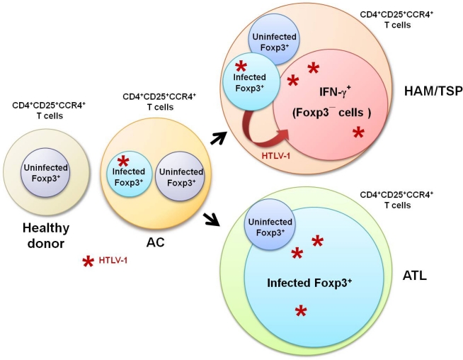
Human T-lymphotropic virus type 1 (HTLV-1) is a retrovirus that is the causative agent of adult T cell leukemia/lymphoma (ATL) and associated with multiorgan inflammatory disorders, including HTLV-1-associated myelopathy/tropical spastic paraparesis (HAM/TSP) and uveitis. HTLV-1-infected T cells have been hypothesized to contribute to the development of these disorders, although the precise mechanisms are not well understood. HTLV-1 primarily infects CD4(+) T helper (Th) cells that play a central role in adaptive immune responses. Based on their functions, patterns of cytokine secretion, and expression of specific transcription factors and chemokine receptors, Th cells that are differentiated from naïve CD4(+) T cells are classified into four major lineages: Th1, Th2, Th17, and T regulatory (Treg) cells. The CD4(+)CD25(+)CCR4(+) T cell population, which consists primarily of suppressive T cell subsets, such as the Treg and Th2 subsets in healthy individuals, is the predominant viral reservoir of HTLV-1 in both ATL and HAM/TSP patients. Interestingly, CD4(+)CD25(+)CCR4(+) T cells become Th1-like cells in HAM/TSP patients, as evidenced by their overproduction of IFN-γ, suggesting that HTLV-1 may intracellularly induce T cell plasticity from Treg to IFN-γ(+) T cells. This review examines the recent research into the association between HTLV-1 and Treg cells that has greatly enhanced understanding of the pathogenic mechanisms underlying immune dysregulation in HTLV-1-associated neuroinflammatory disease.
Keywords: ATL; CD4+CD25+CCR4+ T cell; HAM/TSP; HTLV-1; exFoxp3+ cell; immune-dysfunction; inflammation; regulatory T cell.
Figures


References
-
- Uchiyama T, Yodoi J, Sagawa K, Takatsuki K, Uchino H. Adult T-cell leukemia: Clinical and hematologic features of 16 cases. Blood. 1977;50:481–492. - PubMed
-
- Gessain A, Barin F, Vernant JC, Gout O, Maurs L, Calender A, de The G. Antibodies to human T-lymphotropic virus type-I in patients with tropical spastic paraparesis. Lancet. 1985;2:407–410. - PubMed
-
- Osame M, Usuku K, Izumo S, Ijichi N, Amitani H, Igata A, Matsumoto M, Tara M. HTLV-I associated myelopathy, a new clinical entity. Lancet. 1986;1:1031–1032. - PubMed
-
- Mochizuki M, Watanabe T, Yamaguchi K, Yoshimura K, Nakashima S, Shirao M, Araki S, Takatsuki K, Mori S, Miyata N. Uveitis associated with human T-cell lymphotropic virus type I. Am J Ophthalmol. 1992;114:123–129. - PubMed
Publication types
MeSH terms
Substances
LinkOut - more resources
Full Text Sources
Other Literature Sources
Research Materials
Miscellaneous
