

Case Reports
doi: 10.4061/2011/942360.
Epub 2011 May 31.
A case of a ruptured sclerosing liver hemangioma
Affiliations
- PMID: 21994877
- PMCID: PMC3170855
- DOI: 10.4061/2011/942360
Item in Clipboard
Case Reports
A case of a ruptured sclerosing liver hemangioma
Int J Hepatol.
2011.
Abstract
Hemangiomas are the most common benign tumors found in the liver, typically asymptomatic, solitary, and incidentally discovered. Although vascular in nature, they rarely bleed. We report a case of a 52-year-old woman with a previously stable hemangioma who presented to our hospital with signs and symptoms indicative of spontaneous rupture. We review the literature, focusing on diagnosis and management of liver hemangiomas.
Figures
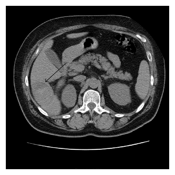
Unenhanced computed tomography of the liver demonstrates a 53.5 mm heterogeneous area in segment six (arrow).
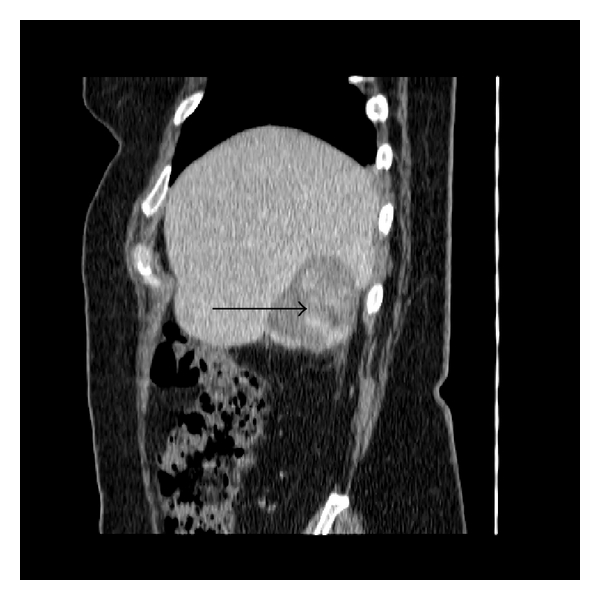
Contrast-enhanced computed tomography of the abdomen in the coronal plane demonstrates intralesional areas of arterial enhancement (arrow). There is a small amount of paracolic gutter fluid.
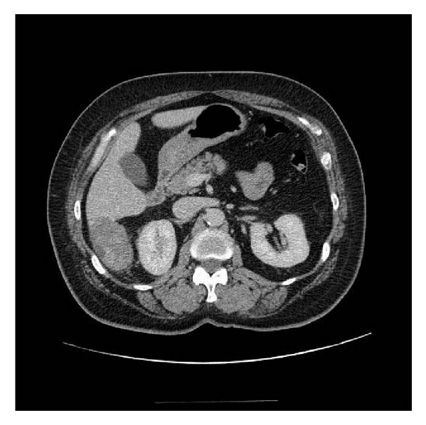
Reimaging of the abdomen with computed tomography three days subsequent to the original study demonstrates further increase in size of the ruptured hemangioma, and it now measures 61.5 mm.
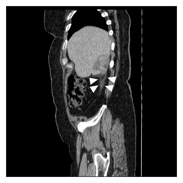
Reimaging of the abdomen with computed tomography three days subsequent to the original study in the coronal plane exhibits an increase in the amount of paracolic gutter fluid (arrowheads), and the craniocaudal dimension of the hemangioma is now 73.8 mm.
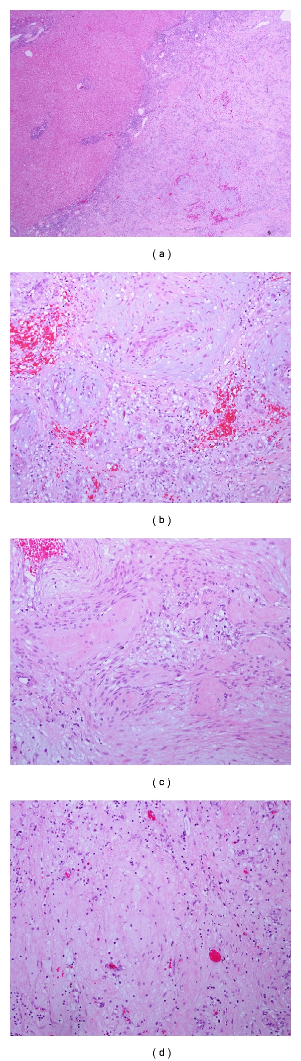
(a) Liver parenchyma with adjacent nonencapsulated mass (H&E, 20x), (b) tumor with a proliferation of thickened blood vessels with myxoid changes (H&E, 400x), (c) the hemangioma shows areas of hyalinized fibrosis (H&E, 400x), and (d) areas of loose connective tissue within the tumor (H&E, 400x).
References
-
- Dietrich CF, Mertens JC, Braden B, Schuessler G, Ott M, Ignee A. Contrast-enhanced ultrasound of histologically proven liver hemangiomas. Hepatology. 2007;45(5):1139–1145. - PubMed
-
- Geschickter CF, Keasbey LE. Tumors of blood vessels. American Journal of Cancer. 1935;25:p. 568.
-
- Sewell JH, Weiss K. Spontaneous rupture of hemangioma of the liver. Archives of surgery. 1961;83:729–733. - PubMed
-
- Caseiro-Alves F, Brito J, Araujo AE, et al. Liver haemangioma: common and uncommon findings and how to improve the differential diagnosis. European Radiology. 2007;17(6):1544–1554. - PubMed
Publication types
LinkOut - more resources
Full Text Sources
