. 2011;1(5):629-49.
Epub 2011 Apr 26.

The complex interplay between autophagy and NF-κB signaling pathways in cancer cells

The complex interplay between autophagy and NF-κB signaling pathways in cancer cells

Aurore Trocoli et al. Am J Cancer Res. 2011.

Abstract

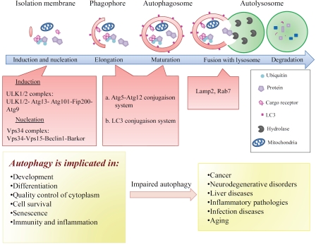
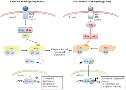
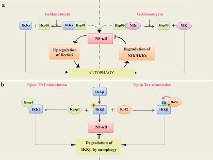
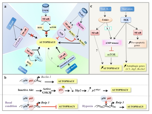
Tight regulation of both the NF-κB pathway and the autophagy process is necessary for maintenance of cellular homeostasis. Deregulation of both pathways is frequently observed in cancer cells and is associated with tumorigenesis and tumor cell resistance to cancer therapies. Autophagy is involved in several cellular functions regulated by NF-κB including cell survival, differentiation, senescence, inflammation, and immunity. On a molecular level, autophagy and NF-κB share common upstream signals and regulators and can control each other through positive or negative feedback loops, thus ensuring homeostatic responses. Here, we summarize and discuss the most recent discoveries that shed new light on the complex interplay between autophagy and NF-κB signaling pathways; this certainly has functional relevance in tumorigenesis and tumor responses to therapy.

Keywords: Autophagy; NF-κB; cancer; signaling pathways.


Figures

Figure 1
Figure 1
Schematic representation of autophagy in mammals. The first step in the induction of autophagy is the formation of a multi-membrane structure, known as a phagophore which further elongates to form an autophagosome before enclosing cytoplasmic material. The autophagosome then fuses with the lysosome to form the autophagoly-sosome, in which the sequestered material is degraded by lysosomal hydrolases. The ULK1/2 complex and the class III phosphatidylinositol 3-kinase (Vps34) complex drive the first step of autophagosome formation (induction and nucleation). The elongation and the closure of the autophagosomal membranes (maturation) require the recruitment of two conjugation systems, Atg5-Atg12 and Atg8/LC3-phosphatidyl ethanolamine, to the phagophore. The lysosomal membrane protein Lamp 2 and the GTPase Rab7 together mediate the fusion of autophagosome with the lysosome. The major cellular functions of autophagy are shown. Depicted are the consequences of impaired autophagy in the development of certain human pathologies. For details, see text.
Figure 2
Figure 2
Schematic model for the activation of NF-κB signaling pathways. In the canonical NF-κB pathway, stimulation of pro-inflammatory receptors activates the IKK complex (comprised of IKKα, IKKβ and IKKγ) leading to the phos-phorylation of IKBCX and its proteasomal degradation. This induces the translocation of p65/p50 dimers into the nucleus where the dimer binds to the promoter regions of a subset of target genes. In the noncanonical NF-κB pathway, stimulation of BAFF-R, CD40, and lymphotoxina-β receptor (LTβR) leads to the stabilization of NIK and results in the activation of the IKKα homo-dimers. Active IKKα, in turn, induces the phosphorylation of the NF-κB family member NF-κB2/p100 leading to its processing by the proteasome to generate p52. This allows the translocation of the p52/Rel B dimer into nucleus where it regulates the transcription of target genes. For details, see text.
Figure 3
Figure 3
Autophagy mediates degradation of NF-κB signaling components. a) NIK and IKKs are targets for degradation by autophagy upon inhibition of Hsp90. This leads to inactivation of NF-κB pathway and, in turn, inhibition of autophagy through downregulation of an essential autophagy gene Beclin 1. The feedback regulatory loop between autophagy and NF-κB pathways is depicted. b) IKKβ degradation by autophagy through a mechanism that involves Keap1. In cells treated by TNF, Keap1 is responsible for the negative regulation of NF-κB through inhibition of the IKKβ phosphorylation and induction of IKKβ degradation by the autophagy pathway. c) IKKβ degradation by autophagy through a mechanism that involves Ro52. In cells infected with HTLV-1, Tax activates IKKβ-dependent NF-κB activation. Active IKKβ subsequently interacts with Ro52; this induces monoubiquitination of IKKβ and leads to its degradation by autophagy resulting in the termination of NF-κB activation. Ub represents ubiquitin. For details, see text.
Figure 4
Figure 4
Regulation of autophagy by NF-κB signaling pathways. a) NF-κB activity regulates the extent of autophagy in cells through NF-κB inducers that regulate autophagy either in a positive or a negative manner. TNF-mediated NF-κB activation represses autophagy through both ROS inhibition and mTOR activation. In macrophages exposed to E. coli, NF-κB activation inhibits autophagy and triggers cell death program. TLR4 stimulation activates autophagy by inducing the ubiquitination (Ub represents ubiquitin) of Beclin 1 by TRAF6, an upstream activator of NF-κB. In turn, NF-κB activation upregulates the expression of A20, a factor that inhibits the ubiquitination of Beclin 1 and thereby limits autophagy. Upon hypoxia, ROS-mediated the activation of both NF-κB and Hif-1α is responsible for the induction of autophagy. In response to heat shock stress, autophagy is induced through a mechanism that involves NF-κB activation, b) NF-κB regulates the transcription of autophagy regulatory genes. In some experimental settings, p65, a member of NF-κB family, upregulates Beclin 1 mRNA level and induces autophagy. In response to DNA damage, the p52 subunit of NF-κB subunit controls autophagy by modulating the expression of both Skp2 and p27KIP1. The p65 sub-unit of NF-κB blocks E2F1-dependent Bnip 3 promoter binding and Bnip 3 transcription, thus inhibiting mitochondrial turnover via autophagy. c) NF-κB signaling components regulate autophagy. TAK1 enhances autophagy through AMP-mediated inhibition of mTORC1. X represents an as-yet-unidentified factor that is required for autophagy induction by TAK1. Under starvation condition, active IKKs induce autophagy through an AMPK inhibition-dependent of mTOR. In starved cells, IKK activity also promotes the upregulation of several essential autophagy genes and a subset of anti-apoptotic genes through an NF-κB independent or dependent mechanism, respectively.

References

    1. He C, Klionsky DJ. Regulation mechanisms and signaling pathways of autophagy. Annu Rev Genet. 2009;43:67–93. - PMC - PubMed
    1. Mizushima N, Levine B. Autophagy in mammalian development and differentiation. Nat Cell Biol. 2010;12:823–830. - PMC - PubMed
    1. Botti J, Djavaheri-Mergny M, Pilatte Y, Codogno P. Autophagy signaling and the cogwheels of cancer. Autophagy. 2006;2:67–73. - PubMed
    1. Ravikumar B, Sarkar S, Davies JE, Futter M, Garcia-Arencibia M, Green-Thompson ZW, Jimenez-Sanchez M, Korolchuk VI, Lichtenberg M, Luo S, Massey DC, Menzies FM, Moreau K, Narayanan U, Renna M, Siddiqi FH, Underwood BR, Winslow AR, Rubinsztein DC. Regulation of mammalian autophagy in physiology and pathophysiology. Physiol Rev. 2010;90:1383–1435. - PubMed
    1. Behrends C, Sowa ME, Gygi SP, Harper JW. Network organization of the human autophagy system. Nature. 2010;466:68–76. - PMC - PubMed

LinkOut - more resources