. 2011;1(5):663-73.
Epub 2011 May 2.

The adaptor proteins p140CAP and p130CAS as molecular hubs in cell migration and invasion of cancer cells

Affiliations

The adaptor proteins p140CAP and p130CAS as molecular hubs in cell migration and invasion of cancer cells

Paola Di Stefano et al. Am J Cancer Res. 2011.

Abstract

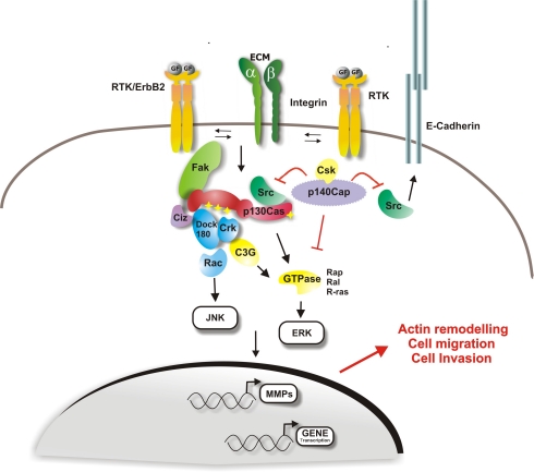
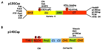
The assembly of molecular hubs upon integrin and growth factor stimulation represents a preferential way to transduce signals throughout the cell. Among the intracellular kinases that are responsive to integrin and growth factor activation, Src Family Kinases (SFKs) are crucial regulators of cell migration and invasion. Increasing evidence highlight the importance of adaptor proteins in these processes, based on their ability to create signalling platforms that control downstream signals. Among these adaptors we will discuss the molecular features of p130Cas and p140Cap proteins in terms of regulation of cell migration and invasion in normal and transformed cells.

Keywords: integrins; invasion; migration; p130Cas; p140Cap; receptor tyrosine kinases.


Figures

Figure 1
Figure 1
p130Cas and p140Cap signalling involved in migration and invasion. Upon extracellular matrix binding or growth factors stimulation, integrins and Receptor Protein Tyrosine Kinases (RPTK) represent the major upstream regulators of p130Cas and p140Cap, mainly through the regulation of Src kinase activity. Once tyrosine phosphory-lated by Src, p130Cas recruits proteins that activate downstream pathways, resulting in actin cytoskeleton reorganization, increased cell motility and migration. p130Cas by acting on metalloproteinases (MMPs) promoter is also required for the invasive program. Upon cell matrix adhesion or mitogen stimulus, p140Cap inhibits Src kinase activity and p130Cas tyrosine phosphorylation and p130Cas/Crk complex formation. As a consequence, the effect of p130Cas on actin cytoskeleton re-organization is impaired and cell migration and invasion are inhibited. Moreover, by inactivating Src, p140Cap also regulates the epidermal growth factor receptor (EGFR) pathway through E-cadherin-dependent inactivation of EGFR signalling. p140Cap by interacting with E-cadherin and EGFR at the cell membrane, immobilizes E-Cadherin at the cell membrane thus preventing cell migration and invasion.
Figure 2
Figure 2
p140Cap and p130Cas structure. A) p130Cas consists of an N-terminal SH3 domain, a substrate domain (SD), a serine rich region (SRR), and a C-terminal domain (CT). The main interactors are indicated. In particular, many proteins associate to the N-terminal domain and the Src family kinases (SFKs) bind the CT domain. The 15 YxxP motifs are phosphorylated by Src family kinases to mediate Crk binding. B) p140Cap consists of an N-terminal tyrosine-rich region (Tyr-rich), an actin binding domain (ABD), a proline rich domain (Pro1), a coil-coiled region (C1-C2), two domains rich in charged amino acids (CH1, CH2) and a C-terminal proline rich domain (Pro2). Src, p130Cas, EB3 and Vinexin bind to the Pro2 domain of p140Cap. The binding regions of Cortactin and Csk have yet to be defined.
Figure 3
Figure 3
p140Cap stabilizes E-Cadherin molecules at the cell membrane. E-cadherin stability and recycling at the cell membrane is crucial for cell-contact growth inhibition and inhibition of cell scatter. Our recent results show that p140Cap stabilizes E-cadherin at the cell membrane through the inhibition of Src kinase. p140Cap over-expressing (p140-oe) and Ctr breast cancer MCF-7 cells were co-transfected with E-Cadherin-GFP and Csk-RNAi. E-Cadherin dynamics were analyzed by fluorescence recovery after photobleaching (FRAP). The Fluorescence recovery curves are shown on the left, and the histogram of the % of immobile E-Cadherin fraction on the right (*p<0.05). The data show that p140 over-expressing cells have a higher amount of the immobile fraction (35%) compared to control cells (20%), confirming that p140Cap stabilizes E-Cadherin molecules at the cell membrane. Moreover, E-cadherin membrane dynamics were restored when Src was rescued by Csk knock-down. siRNA oligos decrease the immobile fraction of p140 over-expressing cells to the level of Ctr cells. The figure is modified from [31].
Figure 4
Figure 4
p140Cap impairs EGF-dependent cell scatter through inhibition of Src activity. Adherens junction dynamics are important for cell scatter. p140Cap by increasing the amount of immobilized E-cadherin at the cell surface, regulates the ability of cells to scatter. Left panels: p140Cap over-expressing (p140-oe) and Ctr breast cancer MCF-7 cells infected with GFP or kinase-negative Csk mutant (Csk-KD) recombinant adenoviruses were grown in small islets, serum deprived for 12 h, untreated (a,d) or treated with 50ng/ml EGF for 12 h (b, c, e, and f), fixed, stained with Diff Quick kit, and photographed at 20x magnification (bar is 10 mm). Right panel: the mean area of the colonies was measured with Metamorph software in 20 random fields on six independent experiments (*p<0.05). The data show that p140Cap impairs the EGF-dependent cell scatter ability (panel e) and that the rescue of Src kinase activity is sufficient to restore cell scatter (panel f). The figure is modified from [31].
Figure 5
Figure 5
p130Cas triggers acina invasion of ErbB2 transformed MCF10 cells. p130Cas over-expressing or Mock ErbB2 transformed MCF10 cells were plated on a Matrigel/collagen 1:1 matrix and left un-stimulated or activated for ErbB2 by treating with the small molecule AP1510. 3D invasive protrusions are present only in p130Cas over-expressing and ErbB2 activated acinar structures. The figure is modified from [57].
Figure 6
Figure 6
3D matrix adhesions in invasive acinar structures of ErbB2 transformed MCF10 cells over-expressing p130Cas. p130Cas over-expressing ErbB2 transformed MCF10 cells were plated on a Matrigel/collagen 1:1 matrix and activated for ErbB2. Confocal images representing 3D matrix adhesions characterizing invasive protrusions of p130Cas over-expressing and ErbB2-activated acini. F-actin (purple, panel a), beta-1 integrin (red, panel b) and fibronectin (green, panel c) staining are shown together with merged images (panel d). Magnifications of the insets are shown in panels e, f, g and h. White arrows in panel h indicate co-localization between beta1 integrin and fibronectin. Scale bars: 25 μm. The figure is modified from [57].
Figure 7
Figure 7
Signaling pathways leading to 3D invasion of ErbB2 transformed MCF10 cells over-expressing p130Cas. PI3K/Akt and Erk1/2 pathways are both activated during invasion triggered by ErbB2 transformation of p130Cas over-expressing mammary epithelial cells. ErbB2/p130Cas/Erk1/2 MAPK signalling pathway preferentially targets mTOR/p70S6K, whereas the ErbB2/p130Cas/PI3K/Akt cascade triggers Rac1 activation. Both signaling pathways are required for mammary epithelia invasion in 3D suggesting that they cooperate in the regulation of different processes that ultimately lead to cell invasion. The figure is modified from [57].

References

    1. Ridley AJ, Schwartz MA, Burridge K, Firtel RA, Ginsberg MH, Borisy G, Parsons JT, Horwitz AR. Cell migration: integrating signals from front to back. Science. 2003;302:1704–1709. - PubMed
    1. Mitra SK, Hanson DA, Schlaepfer DD. Focal adhesion kinase: in command and control of cell motility. Nat Rev Mol Cell Biol. 2005;6:56–68. - PubMed
    1. Eliceiri BP, Puente XS, Hood JD, Stupack DG, Schlaepfer DD, Huang XZ, Sheppard D, Cheresh DA. Src-mediated coupling of focal adhesion kinase to integrin alpha(v)beta5 in vascular endothelial growth factor signaling. J Cell Biol. 2002;157:149–160. - PMC - PubMed
    1. Berx G, Raspe E, Christofori G, Thiery JP, Sleeman JP. Pre-EMTing metastasis? Recapitulation of morphogenetic processes in cancer. Clin Exp Metastasis. 2007;24:587–597. - PubMed
    1. Giancotti FG. A structural view of integrin activation and signaling. Dev Cell. 2003;4:149–151. - PubMed