. 2011 Dec 9;286(49):42141-42149.
doi: 10.1074/jbc.M111.254243. Epub 2011 Oct 12.

Capturing a fusion intermediate of influenza hemagglutinin with a cholesterol-conjugated peptide, a new antiviral strategy for influenza virus

Affiliations

Capturing a fusion intermediate of influenza hemagglutinin with a cholesterol-conjugated peptide, a new antiviral strategy for influenza virus

Kelly K Lee et al. J Biol Chem. .

Abstract

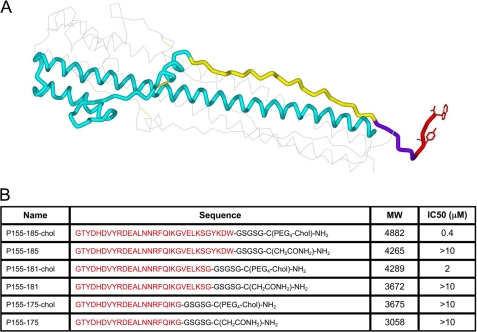
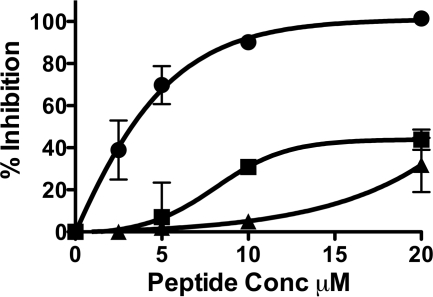
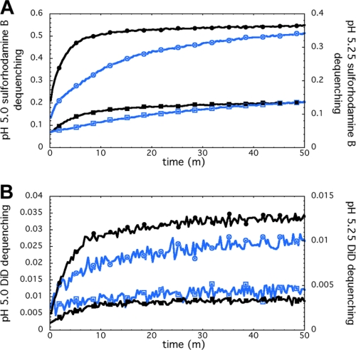
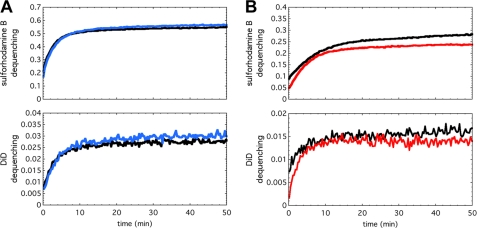
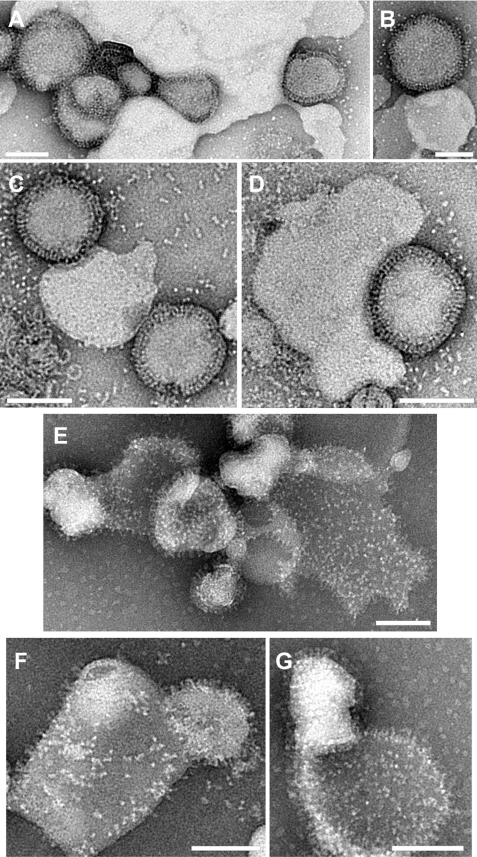
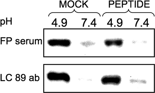
We previously described fusion-inhibitory peptides that are targeted to the cell membrane by cholesterol conjugation and potently inhibit enveloped viruses that fuse at the cell surface, including HIV, parainfluenza, and henipaviruses. However, for viruses that fuse inside of intracellular compartments, fusion-inhibitory peptides have exhibited very low antiviral activity. We propose that for these viruses, too, membrane targeting via cholesterol conjugation may yield potent compounds. Here we compare the activity of fusion-inhibitory peptides derived from the influenza hemagglutinin (HA) and show that although the unconjugated peptides are inactive, the cholesterol-conjugated compounds are effective inhibitors of infectivity and membrane fusion. We hypothesize that the cholesterol moiety, by localizing the peptides to the target cell membrane, allows the peptides to follow the virus to the intracellular site of fusion. The cholesterol-conjugated peptides trap HA in a transient intermediate state after fusion is triggered but before completion of the refolding steps that drive the merging of the viral and cellular membranes. These results provide proof of concept for an antiviral strategy that is applicable to intracellularly fusing viruses, including known and emerging viral pathogens.


Figures

FIGURE 1.
FIGURE 1.
Cholesterol-conjugated peptides derived from influenza HA are effective influenza virus entry inhibitors. A, structure of the influenza virus hemagglutinin in the low-pH post-fusion conformation, highlighting the region corresponding to the cholesterol-conjugated fusion inhibitors. Only one of the three units of the trimer is shown for clarity. The region corresponding to the cholesterol-tagged inhibitors and their controls is highlighted in yellow, violet and red. Yellow, amino acids 155–175, common to all peptides; violet, amino acids 176–181; red, amino acids 182–185. The linker GSGSG and the cholesterol group are C-terminal to the last HA2 residue included in the inhibitor. B, table showing the 6 HA derived peptides and their IC50 versus influenza H3N2 live virus in a plaque reduction assay. The amino acids from the HA sequence are shown in red.
FIGURE 2.
FIGURE 2.
H3 HA derived cholesterol conjugated peptides inhibit 1918 influenza HA mediated infection. Inhibition of viral entry by P155–185-chol (●), P155–185 (■), and HIV-derived cholesterol tagged peptide (▴) using 1918 HA-pseudotyped VSV-ΔG-RFP (vesicular stomatitis virus engineered to express red fluorescent protein and lacking its own G envelope protein, substituted by the 1918 HA).
FIGURE 3.
FIGURE 3.
H3 HA-derived cholesterol conjugated peptides block influenza virus fusion. Fluorescence-monitored assays of fusion and content leakage used cholesterol-tagged inhibitor (P155–185-chol) added directly to preformed liposomes (4:1 w/w DOPC:cholesterol, 200-nm diameter). A water-soluble fluorophore, sulforhodamine B, was encapsulated in the aqueous lumen of the liposomes at self-quenching concentrations. The lipophilic fluorophore, DiD, was embedded in the viral membrane, also at self-quenching concentrations. Inhibitor-decorated liposomes were incubated with X-31 H3N2 virus prior to acidification to pH 5.25 (blue traces) or 5.0 (black traces). A and B, the fluorescence signals increased as a result of fluorophore dilution and fluorescence dequenching. Fluorescence assay at pH 5.25, exhibited inhibitor concentration-dependent inhibition of sulforhodamine B leakage (A) as well as inhibition of lipid mixing (B). 0, 1, 3, and 5 μm inhibitor reactions were performed. Only 0 μm (traces labeled with circles) and 5 μm (traces labeled with squares) data are shown here for clarity. 1 and 3 μm cases were intermediate in the degree of inhibition. pH 5.0 experiments exhibited the same concentration-dependent inhibition trends with faster kinetics.
FIGURE 4.
FIGURE 4.
Fusion inhibition depends on the cholesterol anchor and the specific membrane in which the antiviral peptide is anchored. (A) and (B), fluorescence-monitored fusion reactions with sulforhodamine B fluorescence (encapsulated within the liposomes), which reports liposome permeabilization (top panels), and DiD fluorescence (embedded in the viral membrane), which reports lipid mixing (bottom panels). (A), if P155–185 is not anchored to a membrane by conjugation to cholesterol, inhibition is abolished. P155–185 was incubated at 5 μm (blue traces) with 4:1 w/w DOPC:cholesterol liposomes prior to mixing with virus and acidifying to pH 5.0. The fluorescence-dequenching traces reporting on liposome content leakage (top panel) and lipid mixing (bottom panel) are essentially superimposable with 0 μm inhibitor controls (black traces). (B), P155–185-chol was incubated at 10 μm only with virus prior to mixing with DOPC:cholesterol liposomes and acidification to pH 5.0 to initiate the fusion reaction. Even with 10 μm peptide preincubated with viral particles (red traces), inhibition of liposome leakage (top panel) and lipid mixing (bottom panel) is minimal, and the fluorescence-dequenching traces closely follow the control reactions with no inhibitor present (black traces).
FIGURE 5.
FIGURE 5.
Trapping the HA in a transient intermediate state with cholesterol-conjugated peptides. Negative-stain transmission electron microscopy, comparing liposome-virus complexes with 5 μm inhibitor at pH 5.25 (A and B) and pH 5.5 (C and D). In the presence of the inhibitor, the virus is closely associated with liposomes (suggesting fusion-peptide mediated interaction occurring in the absence of sialic acid influenza receptor present), the membranes have not joined, and there is no evidence of dispersed glycoproteins on liposomes. In the samples without the inhibitor present after incubation at pH 5.25 (E) or pH 5.5 (F and G), the virus and liposomes show significant merging of membranes, viral glycoprotein spikes have dispersed across the liposomal surface, and the liposomes appear flattened and more permeable to stain, suggesting a loss of bilayer integrity. Scale bars = 100 nm.
FIGURE 6.
FIGURE 6.
Cholesterol-conjugated peptides do not prevent pH-induced HA activation. Monolayers of cells expressing HA were incubated for 30 min in media at pH 4.9 or pH 7.4 with or without 50 μm P155–185-chol peptide as indicated. HA conformation was analyzed by immunoprecipitation with the conformation-specific antibodies (ab) LC89 and anti-FP antiserum. Representative Western blots show HA detected by polyclonal anti-GFP-HRP-conjugated antibodies. We note that the fusion peptide sequence recognized by the FP serum is highly conserved across serotypes. The LC89 antibody was originally raised against X-31 HA that had been treated with acidic pH. The HA2 residues recognized by LC89, 106–112, are HTIDLT in X-31. In 1918 H1N1, residues 106–112 are RTLDFH. The sequence specificity of LC89 binding is linked primarily to having a threonine at position 107. If this is mutated, LC89 binding is greatly reduced (38, 39). Given the conservation of Thr-107 in X-31 and H1N1 HA2, the LC89 antibody may be able to still recognize the conformational epitope in our experiments in with HA from 1918 H1N1. In terms of the pattern of ionizable, polar, and apolar residues, the two sequences are also similar.

References

    1. Moscona A. (2008) Annu. Rev. Med. 59, 397–413 - PubMed
    1. Moscona A. (2005) N. Engl. J. Med. 353, 1363–1373 - PubMed
    1. Moscona A. (2009) N. Engl. J. Med. 360, 953–956 - PubMed
    1. Ingallinella P., Bianchi E., Ladwa N. A., Wang Y. J., Hrin R., Veneziano M., Bonelli F., Ketas T. J., Moore J. P., Miller M. D., Pessi A. (2009) Proc. Natl. Acad. Sci. U.S.A. 106, 5801–5806 - PMC - PubMed
    1. Porotto M., Carta P., Deng Y., Kellogg G. E., Whitt M., Lu M., Mungall B. A., Moscona A. (2007) J. Virol. 81, 10567–10574 - PMC - PubMed

Publication types