. 2011 Dec 16;286(50):42911-22.
doi: 10.1074/jbc.M111.311662. Epub 2011 Oct 12.

Protein phosphatase 5 mediates lipid metabolism through reciprocal control of glucocorticoid receptor and peroxisome proliferator-activated receptor-γ (PPARγ)

Affiliations

Protein phosphatase 5 mediates lipid metabolism through reciprocal control of glucocorticoid receptor and peroxisome proliferator-activated receptor-γ (PPARγ)

Terry D Hinds Jr et al. J Biol Chem. .

Abstract

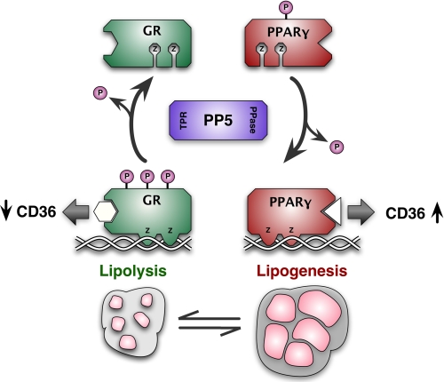
Glucocorticoid receptor-α (GRα) and peroxisome proliferator-activated receptor-γ (PPARγ) regulate adipogenesis by controlling the balance between lipolysis and lipogenesis. Here, we show that protein phosphatase 5 (PP5), a nuclear receptor co-chaperone, reciprocally modulates the lipometabolic activities of GRα and PPARγ. Wild-type and PP5-deficient (KO) mouse embryonic fibroblast cells were used to show binding of PP5 to both GRα and PPARγ. In response to adipogenic stimuli, PP5-KO mouse embryonic fibroblast cells showed almost no lipid accumulation with reduced expression of adipogenic markers (aP2, CD36, and perilipin) and low fatty-acid synthase enzymatic activity. This was completely reversed following reintroduction of PP5. Loss of PP5 increased phosphorylation of GRα at serines 212 and 234 and elevated dexamethasone-induced activity at prolipolytic genes. In contrast, PPARγ in PP5-KO cells was hyperphosphorylated at serine 112 but had reduced rosiglitazone-induced activity at lipogenic genes. Expression of the S112A mutant rescued PPARγ transcriptional activity and lipid accumulation in PP5-KO cells pointing to Ser-112 as an important residue of PP5 action. This work identifies PP5 as a fulcrum point in nuclear receptor control of the lipolysis/lipogenesis equilibrium and as a potential target in the treatment of obesity.


Figures

FIGURE 1.
FIGURE 1.
Interaction of PP5 with GRα and PPARγ. A, Western blot analysis of whole cell extracts from WT and PP5-KO MEF cells demonstrating a complete lack of PP5 in the KO cells and restoration of PP5 expression following rescue of PP5-KO cells with FLAG-PP5 (KO-R). Hsp90 was used as loading control. B, co-immunoadsorption of hormone-free GR heterocomplexes demonstrating preferential interaction with FKBP51 and PP5. GR complexes from WT and PP5-KO cells were adsorbed to protein G-Sepharose using FiGR monoclonal antibody to GR (I) or non-immune IgG (NI) followed by Western blotting for GR and associated TPR proteins. C, co-immunoadsorption of PPARγ with PP5 is dependent on rosiglitazone activation. WT MEF cells were transfected with GFP-PPARγ2 construct followed by a time course of treatment with 1 μm rosiglitazone. Cell extracts were immunoadsorbed with antibody to GFP (I) or non-immune IgG (NI) followed by Western blotting for PPARγ and PP5. A representative of three independent experiments is shown.
FIGURE 2.
FIGURE 2.
PP5 is required for lipogenesis and induction of adipogenic genes. A, detection of lipid accumulation in WT, PP5-KO, and rescued PP5-KO cells by Nile Red staining. Adipogenic differentiation (D) or undifferentiation (U) was achieved by treatment of MEF cells with DIIR mixture for 8 days followed by staining for lipid content. Phase-contrast images are also shown. B, quantification of intracellular lipid in cells treated in A. ###, p < 0.001 (versus WT undifferentiation); ***, p < 0.001 (versus WT differentiation); ∧∧∧, p < 0.001 (versus PP5-KO differentiation) (±S.E.; n = 3). C, biochemical quantification of free fatty acid content in the growth media of cells treated in A. ##, p < 0.01 (versus WT undifferentiation); **, p < 0.01 (versus WT differentiation); #, p < 0.05 (versus WT differentiation) (±S.E.; n = 3). D, fatty-acid synthase (FAS) enzymatic activity of cells treated in A. ##, p < 0.01 (versus WT undifferentiation); ***, p < 0.001 (versus WT differentiation). E, PP5 is required for induction of adipogenic markers. qRT-PCR analysis of WT, PP5-KO, and rescued PP5-KO (KO-R) cells following differentiation (D) or undifferentiation (U) with DIIR mixture. #, versus WT undifferentiation ; *, versus WT differentiation ; ∧, versus KO differentiation (±S.E.; n = 6–9). *, p < 0.05; **, p < 0.01; ***, p < 0.001. The same parameters apply to # and symbols. HSL, hormone-sensitive lipase.
FIGURE 3.
FIGURE 3.
Role of dexamethasone in MEF cell adipogenesis. A, WT and PP5-KO MEF cells were treated with adipogenic mixture containing Dex on the 1st day of treatment (+Dex) or not containing Dex (−Dex) or with mixture containing Dex on each day of treatment (+Dex ET). Lipid accumulation was detected by Nile Red staining. To test for the involvement of PKA signaling, MEF cells were also exposed to H89 PKA inhibitor on each day of DIIR treatment (+H89 ET). B, direct biochemical measurement of intracellular lipid content in cells of A. *, versus WT same condition; #, versus WT +Dex; ∧, versus KO −Dex, ± S.E., n = 3. ## and ∧∧, p < 0.01; ***, p < 0.001.
FIGURE 4.
FIGURE 4.
Reciprocal regulation of GRα and PPARγ activity by PP5. A, Western blot analysis of endogenously expressed GRα and transiently transfected PPARγ2 in WT and PP5-KO cells. B, absence of PP5 increases GRα activity at endogenous metabolic genes. Undifferentiated WT and PP5-KO MEF cells were treated with or without Dex for 2 h followed by qRT-PCR analysis of the indicated genes. #, WT versus WT; *, KO versus WT; , KO versus KO (±S.E.; n = 6–9). C, absence of PP5 decreases PPARγ activity at select metabolic genes. Undifferentiated WT and PP5-KO MEF cells transfected with PPARγ2 were treated with or without rosiglitazone (Rosi) for 2 h followed by qRT-PCR analysis of the indicated genes. #, WT versus WT; *, KO versus WT; , KO versus KO (± S.E.; n = 6–9). *, #, and , p < 0.05; ** and ##, p < 0.01; ***, ###, and , p < 0.001. GILZ, glucocorticoid-inducible leucine zipper; SGK, serum- and glucocorticoid-inducible kinase; PEPCK, phosphoenolpyruvate carboxykinase; Hes1, hairy enhancer of split; FAS, fatty-acid synthase; SREBP, sterol-responsive element-binding protein; PDK4, pyruvate dehydrogenase kinase-4.
FIGURE 5.
FIGURE 5.
PP5 does not regulate intracellular trafficking of GRα and PPARγ. A, localization of PP5 in WT and PP5-KO MEF cells by indirect immunofluorescence. B, localization of GRα-GFP in WT and PP5-KO cells treated with or without Dex for 1 h. C, localization of PPARγ-GFP in WT and PP5-KO cells treated with or without rosiglitazone (Rosi) for 1 h.
FIGURE 6.
FIGURE 6.
PP5 control of GRα and PPARγ phosphorylation. A, PP5 controls GRα phosphorylation at serines 212 and 234. Whole cell extracts of WT and PP5-KO MEF cells treated with or without Dex for 1 h were analyzed by Western blotting with antibodies specific to serines 212, 220, and 234 of mouse GRα. FiGR antibody was used to detect total GRα. Quantitation of GRα bands was performed by infrared spectrophotometry. Phospho-GR (pGR) signals were normalized to total GR at each condition. #, WT versus WT; *, KO versus WT; , KO versus KO (±S.E.; n = 4). B, PP5 controls PPARγ phosphorylation at serine 112. Whole cell extracts of WT and PP5-KO MEF cells transfected with wild-type PPARγ2 or S112A mutant were analyzed by Western blotting with antibody specific to serine 112 or antibody against total PPARγ. Prior to harvest, cells were untreated (C) or treated with tetradecanoylphorbol acetate (TPA) or rosiglitazone (Rosi). Quantitation of PPARγ2 bands was performed by infrared spectrophotometry. Phospho-PPARγ signals were normalized to total PPARγ at each condition. #, WT versus WT; *, KO versus WT (±S.E.; n = 4). C, absence of PP5 does not reduce activity of the S112A-PPARγ2 mutant. WT and PP5-KO MEF cells transfected with wild-type PPARγ2 or S112A mutant were analyzed for rosiglitazone-induced reporter activity using peroxisome proliferator response element (PPRE)-luciferase (Luc) or by a qRT-PCR assay at the indicated endogenous genes. #, WT versus WT; *, KO versus WT (±S.E.; n = 6). D, the S112A-PPARγ2 mutant rescues lipid accumulation in PP5-KO cells. WT and PP5-KO MEF cells were transfected with wild-type PPARγ2 or S112A mutant followed by treatment with DIIR mixture and Nile Red staining for lipid content. Quantitation of lipid content is shown to the right. #, WT versus WT; *, KO versus WT; , KO versus KO (±S.E.; n = 3). E, the GR3A mutant increases lipid in WT but not PP5-KO cells. WT and PP5-KO MEF cells were transfected with the GR3A mutant followed by treatment with DIIR mixture and Nile Red staining for lipid content. Quantitation of lipid content is shown to the right. #, WT versus WT; *, KO versus WT (±S.E.; n = 3). *, p < 0.05; **, p < 0.01; ***, p < 0.001. The same parameters apply to # and ∧ symbols. PEPCK, phosphoenolpyruvate carboxykinase.
FIGURE 7.
FIGURE 7.
PP5 serves as fulcrum in lipogenesis-lipolysis equilibrium by reciprocally modulating GRα and PPARγ. PP5 is a reciprocal modulator of GRα and PPARγ that antagonizes the antilipogenic actions of GRα while simultaneously promoting the lipogenic actions of PPARγ. PP5 achieves this by selectively dephosphorylating GR at serines 212 and 234, causing inhibition of GRα activity at genes, such as CD36. PP5 dephosphorylates PPARγ at serine 112, promoting its transcriptional activity at prolipogenic genes, for example CD36. PPase, phosphatase.

Similar articles

Cited by

References

    1. Vegiopoulos A., Herzig S. (2007) Mol. Cell. Endocrinol. 275, 43–61 - PubMed
    1. Lehrke M., Lazar M. A. (2005) Cell 123, 993–999 - PubMed
    1. Slavin B. G., Ong J. M., Kern P. A. (1994) J. Lipid Res. 35, 1535–1541 - PubMed
    1. Shi X., Shi W., Li Q., Song B., Wan M., Bai S., Cao X. (2003) EMBO Rep. 4, 374–380 - PMC - PubMed
    1. Sugden M. C., Holness M. J. (2006) Arch. Physiol. Biochem. 112, 139–149 - PubMed

Publication types

MeSH terms