. 2011 Oct;34(8):1200-11.
doi: 10.1111/j.1460-9568.2011.07857.x. Epub 2011 Oct 13.

A cis-acting region in the N-methyl-d-aspartate R1 3'-untranslated region interacts with the novel RNA-binding proteins beta subunit of alpha glucosidase II and annexin A2--effect of chronic ethanol exposure in vivo

Affiliations

A cis-acting region in the N-methyl-d-aspartate R1 3'-untranslated region interacts with the novel RNA-binding proteins beta subunit of alpha glucosidase II and annexin A2--effect of chronic ethanol exposure in vivo

Antje Anji et al. Eur J Neurosci. 2011 Oct.

Abstract

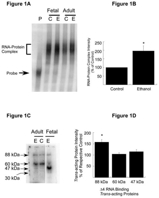
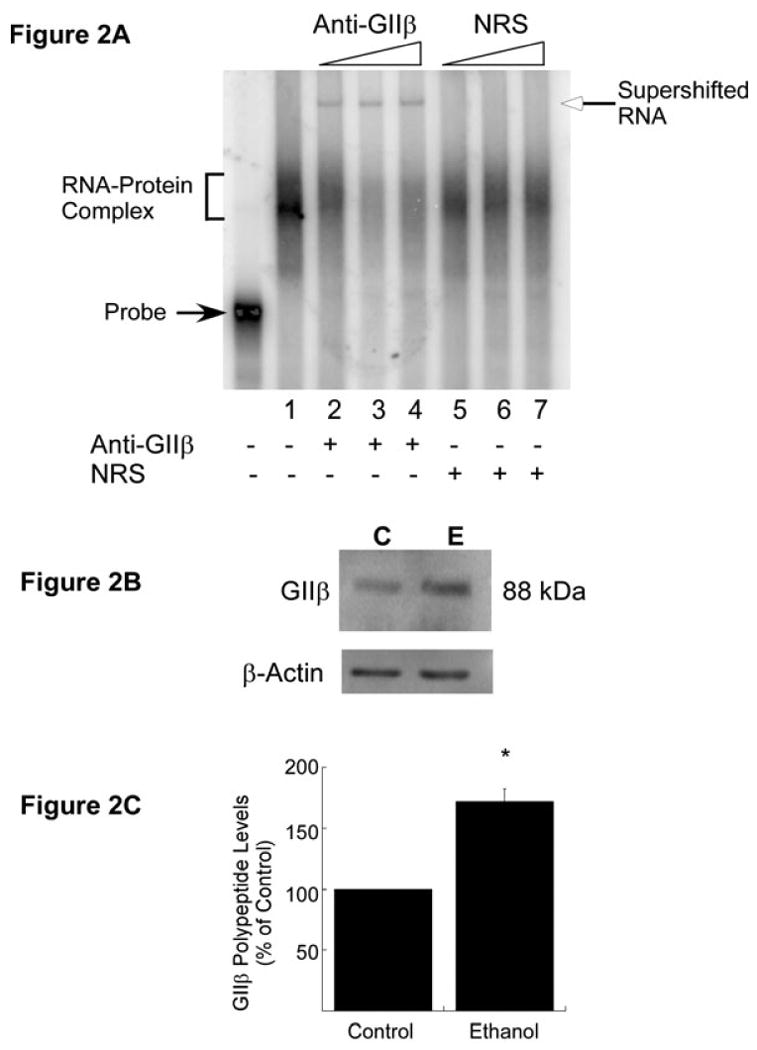
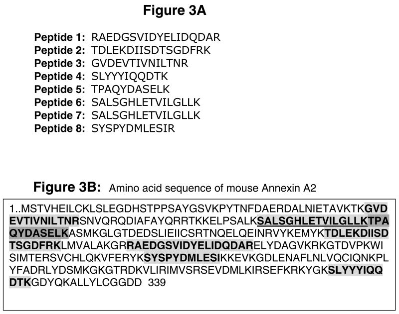
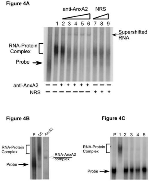
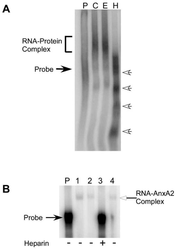
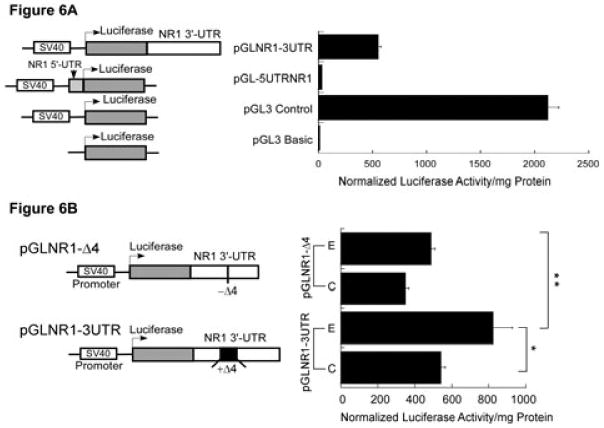
A cis-acting region, Δ4, located in the 3'-untranslated region of N-methyl-d-aspartate R1(NR1) mRNA interacts with several trans-acting proteins present in polysomes purified from fetal cortical neurons. Chronic ethanol exposure of fetal cortical neurons increases Δ4 RNA-protein interactions. This increased interaction is due to an increase in one of the Δ4-binding trans-acting proteins identified as beta subunit of alpha glucosidase II (GIIβ). In this study, we examined whether ethanol-mediated regulation of NR1 mRNA in vivo is similar to that in vitro and whether Δ4-trans interactions are important for ethanol-mediated NR1 mRNA stability. Our data show that polysomal proteins from adult mouse cerebral cortex (CC) formed a complex with Δ4 RNA, suggesting the presence of NR1 mRNA-binding trans-acting proteins in CC polysomes. The intensity of the Δ4 RNA-protein complex was increased with polysomes from chronic ethanol-exposed CC. The Δ4 RNA-protein complex harbored GIIβ and a second trans-acting protein identified as annexin A2 (AnxA2). Ethanol-sensitive GIIβ was upregulated by 70% in ethanol-exposed CC. Heparin, a known binding partner of AnxA2, inhibited Δ4 RNA-protein complex formation. Transient transfection studies using chimeric constructs with and without the Δ4 region revealed that cis-trans interactions are important for ethanol-mediated stability of NR1 mRNA. Furthermore, our data highlight, for the first time, the presence of a binding site on the 3'-untranslated region of NR1 mRNA for AnxA2 and demonstrate the regulation of NR1 mRNA by AnxA2, GIIβ and a third NR1 mRNA-binding protein, which is yet to be identified.


Figures

Fig. 1
Fig. 1
Expression of trans-acting proteins in the CC of adult mouse and effect of chronic ethanol treatment. (A) RNA gel shift analysis. [32P]-labeled Δ4 sense RNA was incubated with polysomes purified from the CC of control (C) and chronic ethanol (E)-exposed adult mice (Adult). Polysomes from control (C) and ethanol-exposed (E) FCNs (Fetal) were included as positive controls. Binding of trans-acting proteins to Δ4 RNA was detected by RNA gel mobility shift assays. A representative autoradiogram is shown here. The Δ4 RNA–protein complex (bracket) and free probe (solid arrow) are indicated on the left. (B) Effect of chronic ethanol on RNA–protein interactions. The intensity of the protein–RNA complex in A was quantitated using ImageQuantTL software. Data are expressed as a percentage of control and plotted values represent the mean + SEM of three independent experiments. Statistical analysis was performed by ANOVA and Scheffe's test (*p < 0.01). (C) Nature of Δ4 RNA-binding trans-acting protein(s) in the CC of adult mice. Polysomal proteins from the CC of control (C) and chronic ethanol-exposed (E) adult mice and ethanol-exposed (E) (50 mM, 5 days) FCNs (Fetal) were separated on SDS–polyacrylamide gel electrophoresis, allowed to refold, blotted onto nitrocellulose membrane and probed with [32P]-labeled Δ4 sense RNA. Three trans-acting proteins that interact with Δ4 RNA were detected in the CC of control and ethanol-exposed mice and four in ethanol-exposed FCNs. Solid arrows on the left indicate the apparent molecular mass of Δ4 RNA-binding trans-acting proteins. Note that gel lanes between Adult and Fetal samples have been omitted. (D) Effect of chronic ethanol on NR1 mRNA-binding trans-acting proteins. Intensities of 88, 60 and 47 kDa protein–Δ4 RNA complexes in C were quantitated using ImageQuantTL software. Data are expressed as a percentage of their respective controls and plotted values represent the mean + SEM of three independent experiments. Statistical analysis was performed by ANOVA and Scheffe's test (*p < 0.01).
Fig. 2
Fig. 2
Trans-acting protein GIIβ in the CC of adult mouse. (A) RNA gel mobility supershift analysis to detect the presence of GIIβ in Δ4 RNA–CC polysomal protein complex. The [32P]-labeled Δ4 sense RNA was first incubated with CC polysomes from ethanol-exposed mice as in RNA gel shift assays. After the addition of increasing concentrations of either anti-GIIβ (lane 1, 0.04 μL; lane 2, 0.0625 μL; lane 3, 0.125 μL; lane 4, 0.25 μL of antiserum) or normal rabbit serum (NRS) (lane 5, 0.0625 μL; lane 6, 0.125 μL; lane 7, 0.25 μL), samples were further incubated at room temperature and processed as in RNA gel shift assays. A representative RNA supershift autoradiogram is shown in A. The Δ4 RNA–protein complex (bracket) and free probe (solid arrow) are indicated on the left. The addition (+) of anti-GIIβ or NRS is indicated below the autoradiogram. An increase in anti-GIIβ or NRS concentration in the reaction mix is indicated by triangles above the autoradiogram. Experiments were repeated three times with similar results. (B) In-vivo effects of chronic ethanol exposure on GIIβ polypeptide levels. CC polysomes from control (C) and ethanol-exposed (E) adult mice were separated by SDS–polyacrylamide gel electrophoresis, blotted onto PVDF membrane and probed with anti-GIIβ. Immunoreactive GIIβ bands were visualized using ECL Plus solution as described in Materials and methods. To control for equal loading, membranes were stripped and reprobed with anti-β-actin. (C) Quantitation of GIIβ polypeptide levels was performed using ImageQuantTL software. The signal intensity of the GIIβ-immunoreactive band was divided by the signal intensity of the β-actin band from the same lane and normalized data are expressed as a percentage of control (mean + SEM of three independent experiments). Statistical analysis was performed by ANOVA and Scheffe's test (*p<0.05).
Fig. 3
Fig. 3
AnxA2 – an NR1 3′-UTR-binding trans-acting protein. A 47 kDa protein band in gel obtained using polysomes from ethanol-exposed CC was processed for in-gel trypsin digestion. Tryptic fragments were analyzed by microcapillary reverse-phase HPLC nano-electrospray tandem mass spectrometry and MS spectra were correlated with known proteins using two different programs. (A) Amino acid sequence of peptides that matched 100% to AnxA2 (gi|18645167). (B) Amino acid sequence of murine AnxA2 (339 residues). Shaded boxes with bold letters indicate the amino acid sequence that matched with eight peptides. Two peptides that spanned the same region of AnxA2 are highlighted by a double underline. Comparison of amino acid sequence of eight matched peptides with AnxA2 demonstrated a 27% sequence coverage.
Fig. 4
Fig. 4
Confirmation of the presence of AnxA2 in Δ4 RNA–protein complex. (A) RNA gel mobility supershift analysis. CC polysomes from ethanol-exposed mice were incubated with 32P-labeled Δ4 sense RNA and then increasing concentrations of either anti-AnxA2 (lane 2, 0.1 μL; lane 3, 0.2 μL; lane 4, 0.3 μL; lane 5, 0.4 μL; lane 6, 0.5 μL) or normal rabbit serum (NRS) (lane 7, 0.3 μL; lane 8, 0.4 μL; lane 9, 0.5 μL) were added. Samples were incubated for an additional 20 min and processed as for RNA gel shift analysis. A representative autoradiogram is shown here. A supershift (arrow on the right) was observed upon addition of anti-AnxA2 (lanes 2-6) but not with normal rabbit serum (lanes 7-9). Free probe and Δ4 RNA–protein complex are indicated on the left. Addition of anti-AnxA2 or normal rabbit serum to the incubation mix is indicated by (+) below the lanes, whereas an increase in the concentration of antibody or normal rabbit serum is indicated by the open triangles above the gel. Experiments were repeated three times with similar results. (B) RNA gel mobility assays. Purified recombinant AnxA2 protein or CC polysomes from control mice were incubated with [32P]-labeled Δ4 sense RNA and samples were processed as detailed in Materials and methods. A shift of Δ4 RNA was observed upon incubation of RNA with CC polysomes (bracket on the left) or full-length recombinant AnxA2 protein (open arrow on the right). Arrow on the left indicates the position of free probe. Note that gel lanes between lane CC and lane AnxA2 have been omitted. Experiments were repeated three times with similar results. (C) Specificity of AnxA2 binding to Δ4 RNA. CC polysomes from control mice (lane 1), increasing concentration of purified recombinant thyroglobulin (lane 2, 0.25 μg protein; lane 3, 0.5 μg protein; lane 4, 1.0 μg protein) and bovine serum albumin (lane 5, 1.0 μg protein) were independently incubated with [32P]-labeled Δ4 sense RNA and samples were processed as detailed in Materials and methods. A shift of Δ4 RNA was observed upon incubation of RNA with CC polysomes (bracket on the left) but not with thyroglobulin or bovine serum albumin. Arrow on the left indicates the position of free probe. Experiments were repeated three times with similar results.
Fig. 5
Fig. 5
Effect of heparin on Δ4 RNA–protein interactions. (A) Polysomes from control (lane C) and ethanol-exposed FCNs (lane E) were incubated with [32P]-labeled Δ4 sense RNA in the absence of heparin and ethanol-exposed CC polysomes were incubated with [32P]-labeled Δ4 sense RNA in the presence of heparin (H). At the end of the incubation, samples were processed for RNA gel shift analysis and results were analyzed on PhosphorImager. A shift of Δ4 RNA is seen in the absence of heparin (bracket on the left). Degradation of Δ4 RNA in the presence of heparin into smaller fragments is indicated by open arrows on the right. Free probe (lane P) is indicated by solid arrow on the left. Experiments were repeated three times with similar results. (B) Purified recombinant AnxA2 protein was incubated with [32P]-labeled Δ4 sense RNA in the absence (lanes 1, 2 and 4) or presence (lane 3) of heparin. Following incubation, samples were processed for RNA gel mobility shift assays. A shift of Δ4 RNA (indicated by open arrow on the right) is observed in the absence (lanes 1, 2 and 4) but not in the presence (lane 3) of heparin. Location of free probe (lane P) is indicated on the left and the addition of heparin to the incubation mix is indicated by (+) below the lanes. Experiments were repeated twice with similar results.
Fig. 6
Fig. 6
Functional analysis of the Δ4 region in the regulation of NR1 mRNA stability. (A) Effect of NR1 UTRs on luciferase enzyme activity. A schematic representation of the structure of chimeric constructs is shown on the left. Transcription (bent arrow) of the luciferase gene is driven by the constitutive SV40 promoter located upstream of the luciferase gene. The 3′-UTR of NR1 (white box) was subcloned in-frame downstream of the luciferase gene, whereas the 5′-UTR of NR1 (gray box) was subcloned in-frame upstream of the luciferase gene. These chimeric plasmids were independently transfected into FCNs and co-transfected with plasmid pRL-TK. Firefly and Renilla luciferase activities were determined by the dual-luciferase reporter assay system. Firefly luciferase activity was normalized and expressed as normalized luciferase enzyme activity per mg protein (histogram on the right). (B) Effect of the Δ4 region on expression of luciferase activity. A schematic representation of the structure of two chimeric constructs, pGLNR1-3UTR (containing the full-length NR1 3′-UTR including the Δ4 region) and pGLNR1-Δ4 (excluding the Δ4 region in the NR1 3′-UTR), is shown on the left. Control and ethanol-exposed FCNs were independently transfected with pGLNR1-3UTR and pGLNR1-Δ4, and co-transfected with pRL-TK. At 2 days after transfection, cell lysates from transfected FCNs were used to determine firefly and Renilla luciferase activities. Firefly luciferase activity was normalized as above. The histogram on the right shows the normalized firefly luciferase activity per mg soluble cellular protein. Data points represent the mean + SEM of three or more experiments. Statistical analysis was performed by ANOVA and Scheffe's test (*p<0.05, **p<0.01).

References

    1. Altschul SF, Madden TL, Schaffer AA, Zhang J, Zhang Z, Miller W, Lipman DJ. Gapped BLAST and PSI-BLAST: a new generation of protein database search programs. Nucleic Acids Res. 1997;25:3389–3402. - PMC - PubMed
    1. Altschul SF, Wootton JC, Gertz EM, Agarwala R, Morgulis A, Schaffer AA, Yu YK. Protein database searches using compositionally adjusted substitution matrices. Febs J. 2005;272:5101–5109. - PMC - PubMed
    1. Anji A, Kumari M. A novel RNA binding protein that interacts with NMDA R1 mRNA: regulation by ethanol. Eur J Neurosci. 2006;23:2339–2350. - PubMed
    1. Anji A, Kumari M. Supplementing the liquid alcohol diet with chow enhances alcohol intake in C57 BL/6 mice. Drug Alcohol Depend. 2008;97:86–93. - PMC - PubMed
    1. Anji A, Shaik KA, Kumari M. Effect of ethanol on lipid-mediated transfection of primary cortical neurons. Ann NY Acad Sci. 2003;993:95–102. discussion 123-104. - PubMed

Publication types

MeSH terms