. 2012 Jan 1;302(1):G77-84.
doi: 10.1152/ajpgi.00301.2011. Epub 2011 Oct 13.

Mechanisms of lysophosphatidylcholine-induced hepatocyte lipoapoptosis

Affiliations

Mechanisms of lysophosphatidylcholine-induced hepatocyte lipoapoptosis

Keisuke Kakisaka et al. Am J Physiol Gastrointest Liver Physiol. .

Abstract

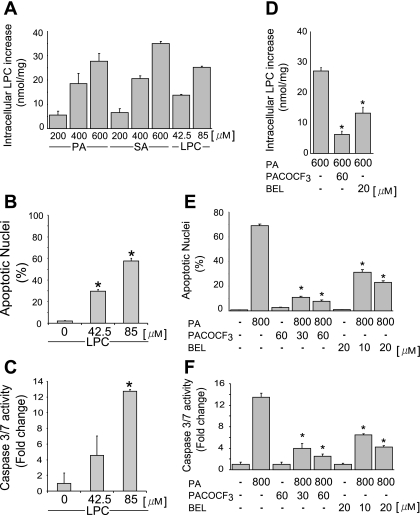
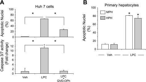
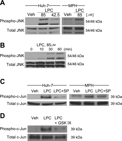
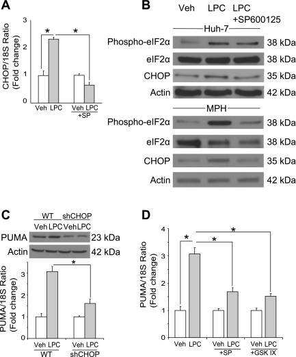
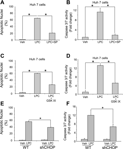
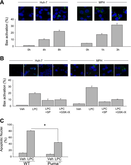
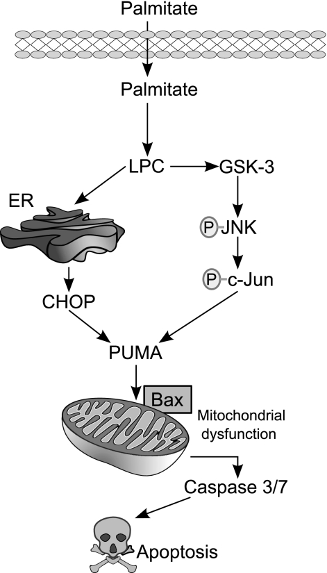
Isolated hepatocytes undergo lipoapoptosis, a feature of hepatic lipotoxicity, on treatment with saturated free fatty acids (FFA) such as palmitate (PA). However, it is unknown if palmitate is directly toxic to hepatocytes or if its toxicity is indirect via the generation of lipid metabolites such as lysophosphatidylcholine (LPC). PA-mediated hepatocyte lipoapoptosis is associated with endoplasmic reticulum (ER) stress, c-Jun NH(2)-terminal kinase (JNK) activation, and a JNK-dependent upregulation of the potent proapoptotic BH3-only protein PUMA (p53 upregulated modulator of apoptosis). Our aim was to determine which of these mechanisms of lipotoxicity are activated by PA-derived LPC. We employed Huh-7 cells and isolated murine and human primary hepatocytes. Intracellular LPC concentrations increase linearly as a function of the exogenous, extracellular PA, stearate, or LPC concentration. Incubation of Huh-7 cells or primary hepatocytes with LPC induced cell death by apoptosis in a concentration-dependent manner. Substituting LPC for PA resulted in caspase-dependent cell death that was accompanied by activating phosphorylation of JNK with c-Jun phosphorylation and an increase in PUMA expression. LPC also induced ER stress as manifest by eIF2α phosphorylation and CAAT/enhancer binding homologous protein (CHOP) induction. LPC cytotoxicity was attenuated by pharmacological inhibition of JNK or glycogen synthase kinase-3 (GSK-3). Similarly, short-hairpin RNA (shRNA)-targeted knockdown of CHOP protected Huh-7 cells against LPC-induced toxicity. The LPC-induced PUMA upregulation was prevented by JNK inhibition or shRNA-targeted knockdown of CHOP. Finally, genetic deficiency of PUMA rendered murine hepatocytes resistant to LPC-induced apoptosis. We concluded that LPC-induced lipoapoptosis is dependent on mechanisms largely indistinguishable from PA. These data suggest that FFA-mediated cytotoxicity is indirect via the generation of the toxic metabolite, LPC.


Figures

Fig. 1.
Fig. 1.
Lysophosphatidylcholine (LPC) is metabolized from saturated fatty acids by PLA2, and inhibition of PLA2 reduces palmitate (PA)-induced apoptosis in hepatocytes. A: intracellular LPC levels were measured using an enzyme-linked colorimetric assay, as described in experimental procedures. The PA-, stearate (SA)-, and LPC-stimulated increase of LPC above basal values is depicted. Huh-7 cells were treated for 12 h with increasing doses of PA, SA (200, 400, and 600 μM) and LPC (42.5 and 85 μM). B and C: Huh-7 cells were treated for 16 h with LPC at either 42.5 or 85 μM, as indicated. Apoptosis was assessed by morphological criteria after 4′,6-diamidine-2′-phenylindole dihydrochloride (DAPI) staining (B). Caspase 3/7 catalytic activity was assessed using a fluorogenic assay and expressed as fold change (C). D: intracellular LPC levels were measured as described in A. Huh-7 cells were treated for 12 h with PA alone or with PA and the PLA2 inhibitor palmityl trifluoromethyl ketone (PACOCF3) or bromoenol lactone (BEL). E and F: Huh-7 cells were treated for 16 h with PA (800 μM) or with PA and the PLA2 inhibitor PACOCF3 (30 or 60 μM) or BEL (10 or 20 μM). Apoptosis was assessed as described in B and E and caspase 3/7 catalytic activity as in C and F, respectively. All data are means ± SE for 3 experiments. *P < 0.01.
Fig. 2.
Fig. 2.
LPC induces caspase-dependent apoptosis in hepatocytes. A: Huh-7 cells were incubated with LPC (85 μM) in the presence of the pan-caspase inhibitor Q-Val-Asp-OPh (QVD-OPh; 10 μM) for 16 h. Vehicle-treated cells (Veh) were used as controls. Apoptotic nuclei were counted after DAPI staining, and caspase 3/7 catalytic activity was measured using a fluorogenic assay. B: mouse primary hepatocytes (MPH) or human primary hepatocytes (HPH) were treated with LPC for 6 or 3 h, respectively. The concentrations of LPC used were 75 μM for MPH and 85 μM for HPH. Apoptosis was assessed as in A. All data are means ± SE for 3 experiments. *P < 0.01.
Fig. 3.
Fig. 3.
LPC induces JNK phosphorylation, and JNK inhibition and glycogen synthase kinase-3 (GSK-3) inhibition reduce c-Jun phosphorylation in hepatocytes. A: immunoblot analysis was performed for phosphorylated (phospho-) and total JNK. Whole cell lysates were prepared from Huh-7 cells incubated with LPC (85 or 42.5 μM) for 8 h or from MPH incubated with LPC (85 μM) for 4 h. B: immunoblot analysis was performed for phosphorylated and total JNK. Whole cell lysates were prepared from Huh-7 cells incubated with LPC (85 μM) for the indicated times. C: immunoblot analysis was performed for phosphorylated and total c-Jun. Whole cell lysates were prepared from Huh-7 cells incubated with LPC at 85 μM in the presence of the JNK inhibitor SP600152 (SP) at 50 μM for 8 h or from MPH incubated with LPC (85 μM) in the presence of SP (50 μM) for 4 h. D: immunoblot analysis was performed for phosphorylated and total c-Jun. Whole cell lysates were prepared from Huh-7 cells incubated with LPC (85 μM) in the presence of the GSK-3 inhibitor GSK-IX (2 μM) for 8 h.
Fig. 4.
Fig. 4.
LPC induces JNK-dependent CAAT/enhancer binding homologous protein (CHOP) expression and CHOP-mediated p53 upregulated modulator of apoptosis (PUMA) expression. A: total RNA was prepared from Huh-7 cells treated with LPC (85 μM) for 8 h. Vehicle-treated cells were used as controls. CHOP mRNA was quantified by real-time PCR, normalized to 18S rRNA, and expressed as fold change over vehicle. B: whole cell lysates were prepared from Huh-7 cells treated with LPC (85 μM) for 8 h or from MPH treated with LPC (85 μM) for 4 h. Immunoblot analysis was performed for phosphorylated eIF2α, total eIF2α, and CHOP. Actin was used as a loading control. C: cells were treated as described in B. Whole cell lysates were prepared 16 h after treatment, and total RNA was extracted 8 h after treatment. Immunoblot analysis was performed for PUMA. Actin was used as a loading control. PUMA mRNA was quantified by real-time PCR, normalized to 18S rRNA, and expressed as fold change over vehicle. D: total RNA was prepared from Huh-7 cells treated with LPC (85 μM) in the presence of the JNK inhibitor SP (50 μM) or the GSK-3 inhibitor GSK-IX (2 μM) for 8 h. PUMA mRNA was quantified by real-time PCR as described above. All data are means ± SE for 3 experiments. *P < 0.01.
Fig. 5.
Fig. 5.
Inhibition of JNK and GSK-3 reduces LPC-induced apoptosis, and CHOP mediates LPC-induced apoptosis. A and B: Huh-7 cells were incubated with LPC (85 μM) in the presence of the JNK inhibitor SP (50 μM) for 16 h. Vehicle-treated cells were used as controls. Apoptosis was assessed using morphological criteria after DAPI staining (A). Caspase 3/7 catalytic activity was assessed using a fluorogenic assay and expressed as fold change (B). C and D: Huh-7 cells were incubated with LPC (85 μM) in the presence of the GSK-3 inhibitor GSK-IX (2 μM) for 16 h. Apoptosis was assessed using morphological criteria after DAPI staining (C). Caspase 3/7 catalytic activity was assessed using a fluorogenic assay and expressed as fold change (D). E and F: Huh-7 (wild type; WT) and Huh-7 cells stably expressing shRNA complementary to CHOP (shCHOP) were incubated with LPC (85 μM) for 16 h. Vehicle-treated cells were used as controls. Apoptotic nuclei were counted after DAPI staining, and caspase 3/7 catalytic activity was measured using a fluorogenic assay. All data are means ± SE for 3 experiments. *P < 0.01.
Fig. 6.
Fig. 6.
LPC-induced Bax activation is JNK and GSK-3 dependent, and LPC-induced apoptosis is mediated by PUMA. A: Huh-7 cells or MPH were incubated with LPC (85 μM) for the indicated times. Representative images of 3 independent experiments are depicted. 6A7-immunoreactive cells, indicative of an activating conformational change in Bax, were quantified in 10 random ×40 objective fields for each condition, and the percentages of 6A7-positive vs. total cells are shown in bar graphs. B: Huh-7 cells or MPH were incubated with LPC (85 μM) in the presence of the JNK inhibitor SP (50 μM) or the GSK-3 inhibitor GSK-IX (2 μM) for 8 or 3 h, respectively. Hepatocytes were analyzed for Bax 6A7 immunoreactivity as described in A. C: MPH were isolated from either WT or Puma−/− mice and treated with LPC (75 μM) for 6 h. Vehicle-treated cells were used as controls. All data are means ± SE for 3 experiments. *P < 0.01.
Fig. 7.
Fig. 7.
Schematic representation of the proposed model for the mechanism of LPC-induced apoptosis. PA-derived LPC induces endoplasmic reticulum (ER) stress and JNK activation, resulting in the induction of the BH3-only protein PUMA. Increased PUMA causes subsequent Bax activation, mitochondrial dysfunction, caspase 3/7 activation, and cellular demise.

References

    1. Adams LA, Lymp JF, Sauver J, Sanderson SO, Lindor KD, Feldstein A, Angulo P. The natural history of nonalcoholic fatty liver disease: a population-based cohort study. Gastroenterology 129: 113–121, 2005 - PubMed
    1. Akazawa Y, Cazanave S, Mott JL, Elmi N, Bronk SF, Kohno S, Charlton MR, Gores GJ. Palmitoleate attenuates palmitate-induced Bim and PUMA up-regulation and hepatocyte lipoapoptosis. J Hepatol 52: 586–593, 2010 - PMC - PubMed
    1. Angulo P, Lindor KD. Non-alcoholic fatty liver disease. J Gastroenterol Hepatol 17, Suppl: S186–S190, 2002 - PubMed
    1. Ariyama Y, Tanaka Y, Shimizu H, Shimomura K, Okada S, Saito T, Yamada E, Oyadomari S, Mori M. The role of CHOP messenger RNA expression in the link between oxidative stress and apoptosis. Metabolism 57: 1625–1635, 2008 - PubMed
    1. Barreyro FJ, Kobayashi S, Bronk SF, Werneburg NW, Malhi H, Gores GJ. Transcriptional regulation of Bim by FoxO3A mediates hepatocyte lipoapoptosis. J Biol Chem 282: 27141–27154, 2007 - PubMed

Publication types

MeSH terms