Comparative Study
. 2011;13(5):R166.
doi: 10.1186/ar3486. Epub 2011 Oct 13.

Increased frequency of circulating Th22 in addition to Th17 and Th2 lymphocytes in systemic sclerosis: association with interstitial lung disease

Affiliations
Comparative Study

Increased frequency of circulating Th22 in addition to Th17 and Th2 lymphocytes in systemic sclerosis: association with interstitial lung disease

Marie-Elise Truchetet et al. Arthritis Res Ther. 2011.

Abstract

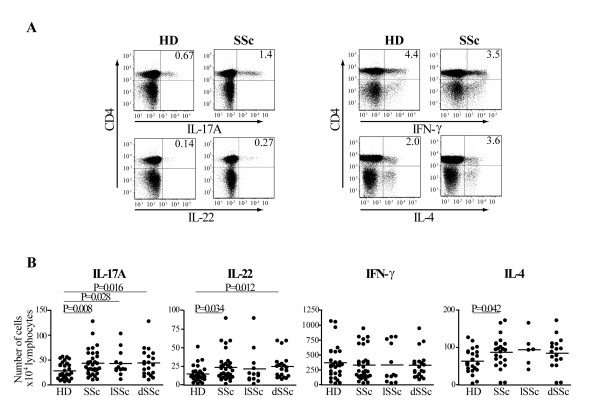
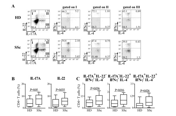
Introduction: T cell abnormalities have been associated with the pathogenesis of systemic sclerosis (SSc). Recently, besides T helper (Th)17 cells, the Th22 subset has been identified in humans. Our purpose was to investigate the pattern of cytokines produced and chemokine-receptors expressed by peripheral blood (PB) Th cells in SSc and healthy donors (HD) focusing on cells producing interleukin (IL)-17 and IL-22 and to identify specific clinical associations.

Methods: Clinical data and peripheral blood were collected in 33 SSc individuals and 29 HD. IL-17A, IL-22, interferon gamma (IFN-γ), IL-4 production, the chemokine receptors CCR4, CCR6, CCR10, CXCR3 expression and the CD161 Th17 cell marker were assessed by multiparametric flow cytometry in PB CD4+ T cells. Intracellular cytokine accumulation was further investigated in CD4+ T cells expanded in vitro for seven days.

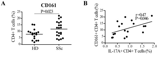
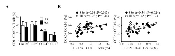
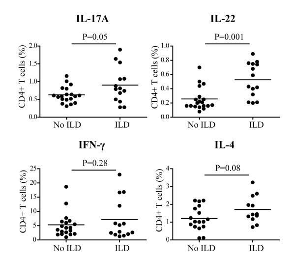
Results: The frequency of Th22, Th17, Th2, but not Th1 cells, was significantly increased in SSc individuals compared to HD. The percentage of CD161+CD4+ T cells was increased in SSc and correlated with the percentage of IL-17A producing cells. Moreover, the expression of the skin- and lung-homing chemokine receptor CCR6 correlated with the frequency of IL-22 and IL-17A-producing cells in SSc but not in HD. Finally, SSc interstitial lung disease (ILD) was strongly associated with higher numbers of IL-22 and, to a lesser extent, IL-17A-producing cells.

Conclusions: IL-22 and IL-17A-producing T cells with skin- and lung-homing capabilities are characteristically increased in SSc. These findings support the hypothesis that Th22, in addition to Th17 cells, may be involved in pathological processes leading to SSc. While the association between IL-22 producing cells and ILD needs to be assessed in larger cohorts of patients, the increased frequency of Th22 cells appears to be a useful novel biomarker in SSc.


Figures

Figure 1
Figure 1
The number of IL-17A+ and IL-22+ CD4+ T cells is increased in SSc at Day 0. PBMC were activated by CD3/CD28 crosslinking for 24 h and surface/intracellular stained for FACS analysis. A. FACS plots gated on CD4+ T cells for representative healthy donors (HD) and SSc individuals. Numbers in plots indicate the percentage of IL-17A, IL-22, IFN-γ and IL-4 producing CD4+ T cells. B. Numbers of IL-17A, IL-22, IFN-γ and IL-4 positive CD4+ T cells for 104 living lymphocytes in 29 HD and 33 SSc (IL-17A, IL-22, IFN-γ) or 22 HD and 23 SSc (IL-4) individuals. Shown are significant differences assessed by unpaired t-test. lSSc, limited cutaneous SSc and dSSc, diffuse cutaneous SSc.
Figure 2
Figure 2
Th17 and Th22 cells are preferentially expanded in SSc individuals. PBMC were activated by CD3/CD28 crosslinking and cultured in the presence of IL-2 (20 U/ml). A. FACS plots gated on CD4+ T cells of cells harvested at Day 7 of culture and activated by PMA/ionomycin for a representative HD and SSc individual. Intracellular cytokine accumulation was detected by five-color flow cytometry. Numbers in plots indicate the percentage of cells in each quadrant. IFN-γ and IL-4 double negative cells are highlighted by grey shading. B. Frequency of IL-17A+ and IL-22+ CD4+ T cells in 29 HD and 30 SSc individuals detected by flow cytometry. C. Frequency of CD4+ T cells producing IL-17A alone (IL-17A+IL-22-IFN-γ-IL-4-cells), IL-22 alone (IL-17A-IL-22-IFN-γ-IL-4-cells), and IL-17A in combination with IL-22 (IL-17A-IL-22-IFN-γ-IL-4-cells) in 24 HD and 29 SSc individuals detected by multiparameter flow cytometry analysis.
Figure 3
Figure 3
CD161+ CD4+ T cells are increased in SSc and correlated with the percentage of IL-17A+CD4+ T cells. A, CD4+CD161+ T cells in the peripheral blood of SSc individuals (n = 21) and HD (n = 17) were identified by two-color flow-cytometry analysis. Bars show the means. B, Correlation between the percentage of IL-17A+CD4+ T cells and CD161+CD4+ T cells analyzed ex vivo. For cytokine intracellular localization, the conditions were as described in the legend of Figure 2. A two-tailed unpaired t-test (A) and Pearson correlation (B) were used for statistical analysis.
Figure 4
Figure 4
IL-17A and IL-22 production correlates with CCR6+CCR10- expression in SSc CD4+ T cells. A. Frequency of CD4+CD45RA- T cells expressing CXCR3, CCR6, CCR4 or CCR10 in SSc and in HD detected by a six-color flow cytometry. Columns represent mean ± SD. B. Correlation between the percentages of IL-17+ CD4+ T cells or IL-22+ CD4+ T cells with CCR6+CCR10- in CD4+CD45RA- T cells. Full and empty dots represent SSc and HD, respectively. Continuous and dotted regression lines correspond to SSc and HD, respectively. Pearson correlation was used for statistical analysis.
Figure 5
Figure 5
Increased frequency of IL-22 producing cells in SSc individuals presenting with interstitial lung disease. Frequency of IL-17A, IL-22, IFN-γ and IL-4 producing CD4+ T cells in SSc individuals presenting or not with ILD. A two-tailed, unpaired t-test was used for statistical analysis.

Similar articles

Cited by

References

    1. Gabrielli A, Avvedimento EV, Krieg T. Scleroderma. N Eng J Med. 2009;360:1989–2003. doi: 10.1056/NEJMra0806188. - DOI - PubMed
    1. Chizzolini C. Update on pathophysiology of scleroderma with special reference to immunoinflammatory events. Ann Med. 2007;39:42–53. doi: 10.1080/07853890601098152. - DOI - PubMed
    1. Allanore Y, Dieude P, Boileau C. Genetic background of systemic sclerosis: autoimmune genes take centre stage. Rheumatology (Oxford) 2010;49:203–210. doi: 10.1093/rheumatology/kep368. - DOI - PubMed
    1. Prescott RJ, Freemont AJ, Jones CJ, Hoyland J, Fielding P. Sequential dermal microvascular and perivascular changes in the development of scleroderma. J Pathol. 1992;166:255–263. doi: 10.1002/path.1711660307. - DOI - PubMed
    1. Kalogerou A, Gelou E, Mountantonakis S, Settas L, Zafiriou E, Sakkas L. Early T cell activation in the skin from patients with systemic sclerosis. Ann Rheum Dis. 2005;64:1233–1235. doi: 10.1136/ard.2004.027094. - DOI - PMC - PubMed

Publication types

MeSH terms