. 2011 Dec;128(6):1216-1224.e11.
doi: 10.1016/j.jaci.2011.08.035. Epub 2011 Oct 11.

Polyinosinic:polycytidylic acid induces protein kinase D-dependent disassembly of apical junctions and barrier dysfunction in airway epithelial cells

Affiliations

Polyinosinic:polycytidylic acid induces protein kinase D-dependent disassembly of apical junctions and barrier dysfunction in airway epithelial cells

Fariba Rezaee et al. J Allergy Clin Immunol. 2011 Dec.

Abstract

Background: Disruption of the epithelial barrier might be a risk factor for allergen sensitization and asthma. Viral respiratory tract infections are strongly associated with asthma exacerbation, but the effects of respiratory viruses on airway epithelial barrier function are not well understood. Many viruses generate double-stranded RNA, which can lead to airway inflammation and initiate an antiviral immune response.

Objectives: We investigated the effects of the synthetic double-stranded RNA polyinosinic:polycytidylic acid (polyI:C) on the structure and function of the airway epithelial barrier in vitro.

Methods: 16HBE14o- human bronchial epithelial cells and primary airway epithelial cells at an air-liquid interface were grown to confluence on Transwell inserts and exposed to polyI:C. We studied epithelial barrier function by measuring transepithelial electrical resistance and paracellular flux of fluorescent markers and structure of epithelial apical junctions by means of immunofluorescence microscopy.

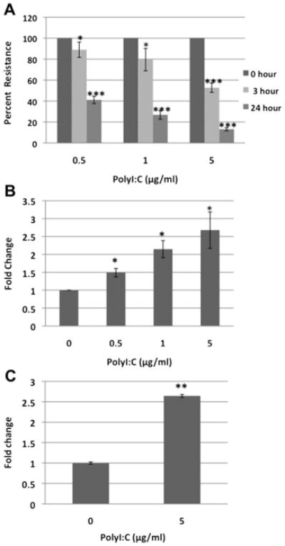
Results: PolyI:C induced a profound decrease in transepithelial electrical resistance and increase in paracellular permeability. Immunofluorescence microscopy revealed markedly reduced junctional localization of zonula occludens-1, occludin, E-cadherin, β-catenin, and disorganization of junction-associated actin filaments. PolyI:C induced protein kinase D (PKD) phosphorylation, and a PKD antagonist attenuated polyI:C-induced disassembly of apical junctions and barrier dysfunction.

Conclusions: PolyI:C has a powerful and previously unsuspected disruptive effect on the airway epithelial barrier. PolyI:C-dependent barrier disruption is mediated by disassembly of epithelial apical junctions, which is dependent on PKD signaling. These findings suggest a new mechanism potentially underlying the associations between viral respiratory tract infections, airway inflammation, and allergen sensitization.


Conflict of interest statement

Disclosure of potential conflict of interest: L. A. Beck receives research support from Centocor, Regeneron, and Genentech. S. N. Georas receives research support from the National Institutes of Health. The rest of the authors declare that they have no relevant conflicts of interest.

Figures

Fig 1
Fig 1
PolyI:C decreases the TEER of our model's airway epithelial cell monolayers. 16HBE14o- cells were grown on Transwell inserts and stimulated with indicated concentrations of Pam3Cys (A), polyI:C (B), LPS (C), flagellin (D), and CpG oligonucleotides (E) for different time periods, followed by TEER measurements. Data are expressed as a percentage of control unstimulated cells and are means ± SEMs of 3 independent experiments per time point. ***P < .001, as determined by using ANOVA, followed by the Student t test with the Bonferonni correction for multiple comparisons.
Fig 2
Fig 2
PolyI:C induces a dose-dependent increase in paracellular permeability. A, TEER was measured in 16HBE14o- cell monolayers stimulated for different times with different concentrations of polyI:C. B and C, Transepithelial flux of sodium fluorescein (Fig 2, B) or 3-kd fluorescein–conjugated dextran (Fig 2, C) was measured after 24 hours of incubation with polyI:C. Data are means ± SEMs of at least 6 independent experiments per group. *P < .05, **P < .01, and ***P < .001, as determined by using the Student t test.
Fig 3
Fig 3
PolyI:C induces disassembly of AJs and TJs. Confluent 16HBE14o-cell monolayers were treated for 6 hours with either medium control or polyI:C (5 μg/mL). Localization of AJ proteins (E-cadherin and β-catenin) and TJ proteins (occludin and ZO-1) was determined by means of immunofluorescence labeling and confocal microscopy. Note the localization of junctional proteins at cell-cell contacts in control cells (arrows) and their translocation into the cytosolic compartment after polyI:C exposure (arrowheads). Images are a representative of at least 6 independent experiments.
Fig 4
Fig 4
Junctional disassembly in polyI:C-exposed epithelial cells is accompanied by remodeling of the apical actin cytoskeleton. Confluent 16HBE14o- cells were exposed for 24 hours to polyI:C (5 μg/mL), followed by fixation and dual-fluorescence labeling for F-actin and ZO-1. PolyI:C induced transformation of the perijunctional F-actin belt (arrows) into a disordered array of apical F-actin bundles (arrowheads). Images are a representative of at least 3 independent experiments.
Fig 5
Fig 5
Effect of PKC isoform inhibitors on polyI:C (pI:C)–induced junctional disassembly. 16HBE14o- cell monolayers were stimulated with polyI:C (5 μg/mL for 24 hours) with or without Gö6976 or Gö6983 (10 μmol/L). Localization of AJ and TJ proteins and architecture of the apical actin cytoskeleton were determined by means of fluorescence labeling and confocal microscopy. Note the normal localization of junctional proteins (arrows) and intact perijunctional F-actin belt (arrowhead) in polyI:C–exposed cells treated with the PKCμ (PKD) inhibitor Gö6976. Images are a representative of at least 3 independent experiments.
Fig 6
Fig 6
Pharmacologic inhibition of PKD attenuates polyI:C-induced epithelial permeability. Control and polyI:C-activated 16HBE14o- cell monolayers (5 μg/mL for 24 hours) were incubated with or without the inhibitor of classical PKC isoforms Gö6976 (10 μmol/L), and permeability was examined by measuring transmonolayer sodium fluorescein flux. Results are expressed as the fold change above control values (increasing values indicating greater permeability) and are presented as means ± SEMs of 3 independent experiments.
Fig 7
Fig 7
PolyI:C-induced activation of PKD in epithelial cells. Confluent 16HBE14o- cell monolayers were treated for 0 to 120 minutes with either medium control or polyI:C (5 μg/mL). Expression of phospho-PKD and total PKD was examined by using Western blotting of whole-cell lysates. Images are representative of 3 independent experiments. PMA, Phorbol 12-myristate 13-acetate.

Comment in

  • Plasticity of airway epithelial cells.
    Avila PC. Avila PC. J Allergy Clin Immunol. 2011 Dec;128(6):1225-6. doi: 10.1016/j.jaci.2011.10.006. J Allergy Clin Immunol. 2011. PMID: 22133319 Free PMC article. No abstract available.

References

    1. Tunggal JA, Helfrich I, Schmitz A, Schwarz H, Gunzel D, Fromm M, et al. E-cadherin is essential for in vivo epidermal barrier function by regulating tight junctions. EMBO J. 2005;24:1146–56. - PMC - PubMed
    1. Niessen CM, Gottardi CJ. Molecular components of the adherens junction. Biochim Biophys Acta. 2008;1778:562–71. - PMC - PubMed
    1. Anderson JM, Van Itallie CM. Physiology and function of the tight junction. Cold Spring Harb Perspect Biol. 2009;1:a002584. - PMC - PubMed
    1. Shen L, Weber CR, Raleigh DR, Yu D, Turner JR. Tight junction pore and leak pathways: a dynamic duo. Annu Rev Physiol. 2011;73:283–309. - PMC - PubMed
    1. Niessen CM. Tight junctions/adherens junctions: basic structure and function. J Invest Dermatol. 2007;127:2525–32. - PubMed

Publication types

MeSH terms