. 2012 Jun;34(5):605-16.
doi: 10.1016/j.medengphy.2011.09.008. Epub 2011 Oct 12.

Three-dimensional finite element analysis of Eustachian tube function under normal and pathological conditions

Affiliations

Three-dimensional finite element analysis of Eustachian tube function under normal and pathological conditions

F J Sheer et al. Med Eng Phys. 2012 Jun.

Abstract

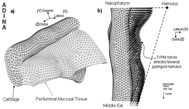
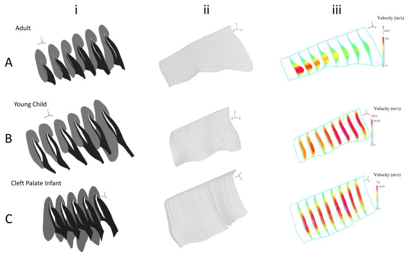
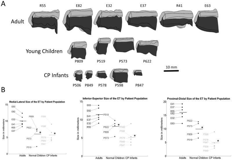
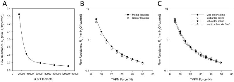
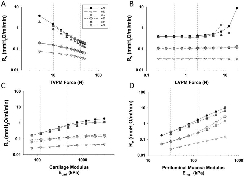
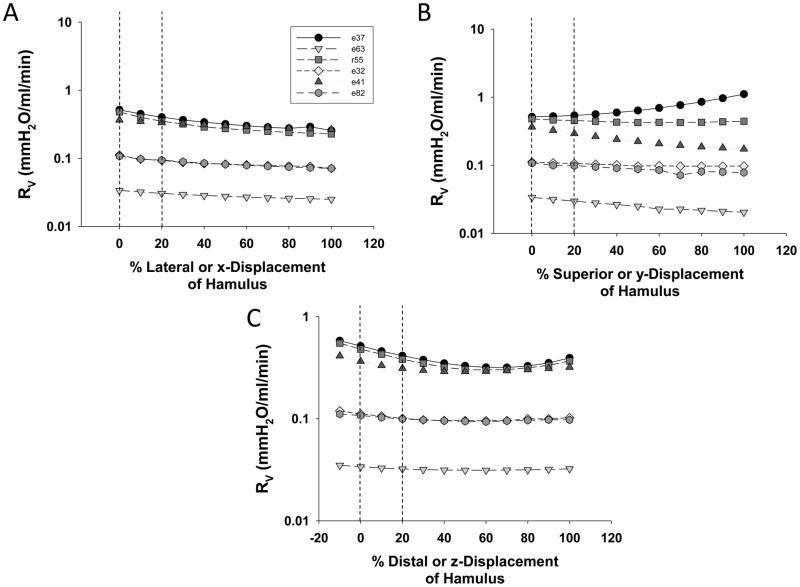
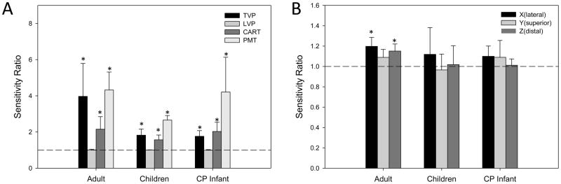
A primary etiological factor underlying chronic middle ear disease is an inability to open the collapsible Eustachian tube (ET). However, the structure-function relationships responsible for ET dysfunction in patient populations at risk for developing otitis media (OM) are not known. In this study, three-dimensional (3D) finite element (FE) modeling techniques were used to investigate how changes in biomechanical and anatomical properties influence opening phenomena in three populations: normal adults, young children and infants with cleft palate. Histological data was used to create anatomically accurate models and FE techniques were used to simulate tissue deformation and ET opening. Lumen dilation was quantified using a computational fluid dynamic (CFD) technique and a sensitivity analysis was performed to ascertain the relative importance of the different anatomical and tissue mechanical properties. Results for adults suggest that ET function is highly sensitive to tensor veli palatini muscle (TVPM) forces and to periluminal mucosal tissue (PMT) elasticity. Young children and cleft palate subjects exhibited reduced sensitivity to TVPM forces while changes in PMT stiffness continued to have a significant impact on ET function. These results suggest that reducing PMT stiffness might be an effective way to restore ET function in these populations. Varying TVPM force vector relationships via changes in hamulus location had no effect on ET opening in young children and cleft palate subjects but did alter force transmission to the ET lumen during conditions of elevated adhesion. These models have therefore provided important new insights into the biomechanical mechanisms responsible for ET dysfunction.


Figures

Figure 1
Figure 1
Serial histological sections of the ET at cross-sectional locations near the middle ear (column i), in the middle of the ET (column ii) and near the nasopharynx (column iii). Images were obtained from healthy adults (top row), young children (middle row) and infants with cleft palate (bottom row). Outlines used to generate 3D finite element models are also shown.
Figure 2AB
Figure 2AB
a) 3D FE model of the ET for a healthy adult subject. b) Superior-Inferior view of the of the TVPM forces directed towards the pytergoid hamulus under baseline conditions.
Figure 3ABC
Figure 3ABC
Slice plots of the deformed solid geometry depicting lumen opening (column i). Grey represents ET cartilage and black represents the mucosal tissue. Finite element meshes within the open lumen used for CFD analysis (column ii). Velocity field within the lumen after simulating pressure driven flow (column iii). Data is shown for one representative subject in each population (a: healthy adults, b: Young children and c: Cleft Palate Infant).
Figure 4AB
Figure 4AB
(A) Solid models created from histological data in all three populations. All models are shown to the same scale and grey represents ET cartilage while black represents the mucosal tissue. (B) Morphological measurements of medial-lateral, inferior-superior and proximal-distal dimensions in each model for the adult, young children and CP infant groups. Horizontal line indicates mean and * indicates differences with respect to (p<0.05).
Figure 5ABC
Figure 5ABC
Data related to model validation. a) Effect of mesh density/number in CFD simulations on the calculation of Rv. b) Effect of closed lumen line location on Rv vs. FTVP relationship. c) Effect of 3D reconstruction algorithm on Rv vs. FTVP relationship.
Figure 6ABCD
Figure 6ABCD
Variation of flow resistance (Rv) as a function of a) TVPM forces, b) LVPM forces, c) cartilage stiffness and d) PMT stiffness in the normal adult population. All parameter studies were conducted by varying the selected parameter and holding the other parameters at baseline values. Dashed lines represent upper and lower limits for calculation of the sensitivity ratio reported in Figure 8.
Figure 7ABC
Figure 7ABC
Variation of flow resistance (Rv) as a function of a) lateral, b) superior and c) distal hamulus position in the normal adult population. Hamulus location is reported as a percentage of the maximal size of the ET in the selected direction. Dashed lines represent upper and lower limits for calculation of the sensitivity ratio reported in Figure 8.
Figure 8AB
Figure 8AB
Mean sensitivity values for a) tissue mechanical properties (TVP forces, LVP forces, cartilage stiffness and PMT stiffness) and b) x, y, z hamulus location in the adult, young children and cleft palate populations. All data is reported as mean ± 95% confidence interval. * indicates statistically significant differences (p<0.05) with respect to unity (dashed line).
Figure 9ABC
Figure 9ABC
Von Misses stress generated at the lumen’s interior surface as a function of hamulus position. Data collected in a model with rigid links to simulate very high adhesion with no ET opening.

Similar articles

Cited by

References

    1. Rovers MM. The burden of otitis media. Vaccine. 2008;26 (Suppl 7):G2–4. - PubMed
    1. Bondy J, et al. Direct expenditures related to otitis media diagnoses: extrapolations from a pediatric medicaid cohort. Pediatrics. 2000;105:E72. - PubMed
    1. Bluestone CD. Eustachian Tube Structure, Function, Role in Otitis Media. BC Decker Inc; Hamilton, Ontario: 2005.
    1. Doyle WJ. Middle Ear Pressure Regulation. In: Rosowski JJ, Merchant SN, editors. The Function and Mechanics of Normal, Diseased and Reconstructed Middle Ears. Kugler Publications; The Hague, Netherlands: 2000. pp. 3–21.
    1. Gaihede M, et al. Middle ear pressure regulation--complementary active actions of the mastoid and the Eustachian tube. Otol Neurotol. 2010;31:603–11. - PubMed

Publication types

MeSH terms