. 2012 Jan 31;375(1-2):100-10.
doi: 10.1016/j.jim.2011.09.013. Epub 2011 Oct 2.

Leukocyte compartments in the mouse lung: distinguishing between marginated, interstitial, and alveolar cells in response to injury

Affiliations

Leukocyte compartments in the mouse lung: distinguishing between marginated, interstitial, and alveolar cells in response to injury

Kathryn E Barletta et al. J Immunol Methods. .

Abstract

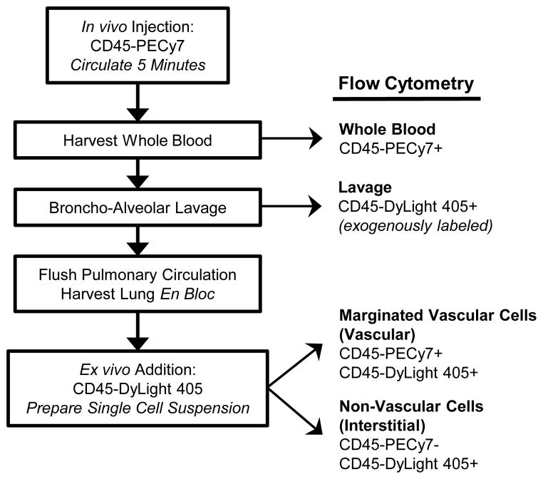
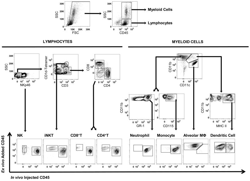
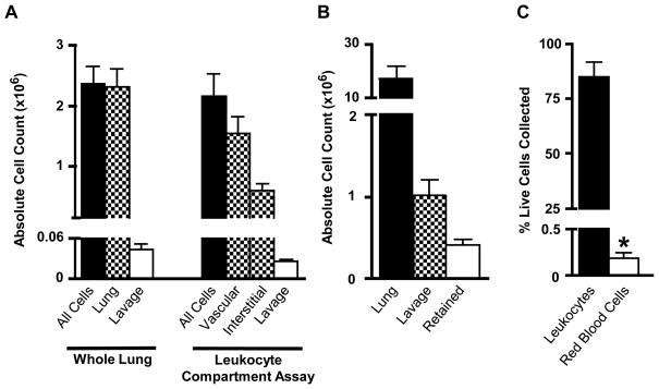
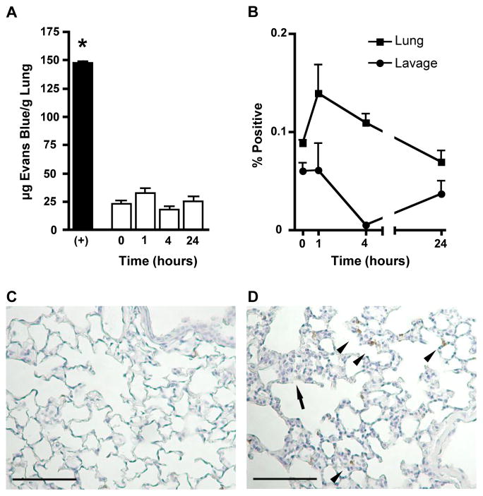
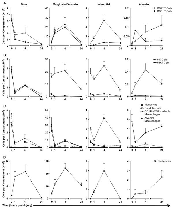
We developed a flow cytometry-based assay to simultaneously quantify multiple leukocyte populations in the marginated vascular, interstitial, and alveolar compartments of the mouse lung. An intravenous injection of a fluorescently labeled anti-CD45 antibody was used to label circulating and marginated vascular leukocytes. Following vascular flushing to remove non-adherent cells and collection of broncho-alveolar lavage (BAL) fluid, lungs were digested and a second fluorescent anti-CD45 antibody was added ex vivo to identify cells not located in the vascular space. In the naïve mouse lung, we found about 11 million CD45+ leukocytes, of which 87% (9.5 million) were in the vascular marginated compartment, consisting of 17% NK cells, 17% neutrophils, 57% mononuclear myeloid cells (monocytes, macrophage precursors and dendritic cells), and 10% T cells (CD4+, CD8+, and invariant NKT cells). Non-vascular compartments including the interstitial compartment contained 7.7×10(5)cells, consisting of 49% NK cells, 25% dendritic cells, and 16% other mononuclear myeloid cells. The alveolar compartment was overwhelmingly populated by macrophages (5.63×10(5)cells, or 93%). We next studied leukocyte margination and extravasation into the lung following acid injury, a model of gastric aspiration. At 1 h after injury, neutrophils were markedly elevated in the blood while all other circulating leukocytes declined by an average of 79%. At 4 h after injury, there was a peak in the numbers of marginated neutrophils, NK cells, CD4+ and CD8+ T cells and a peak in the number of alveolar NK cells. Most interstitial cells consisted of DCs, neutrophils, and CD4+ T cells, and most alveolar compartment cells consisted of macrophages, neutrophils, and NK cells. At 24 h after injury, there was a decline in the number of all marginated and interstitial leukocytes and a peak in alveolar neutrophils. In sum, we have developed a novel assay to study leukocyte margination and trafficking following pulmonary inflammation and show that marginated cells comprise a large fraction of lung leukocytes that increases shortly after lung injury. This assay may be of interest in future studies to determine if leukocytes become activated upon adherence to the endothelium, and have properties that distinguish them from interstitial and circulating cells.


Figures

Fig 1
Fig 1
Leukocyte compartment assay schematic.
Fig 2
Fig 2
Gating strategy for leukocyte compartment assay. Cells were dispersed from mouse lungs, and single cells (singlets, low pulse width) were divided into lymphoid (low SSC) and myeloid (high side scatter, SSC) sub-populations. Lymphoid cells were sub-classified as NK cells (CD3ε NKp46+), invariant NKT cells (CD3ε+ CD1d-tetramer+), CD8+ T cells (CD3ε+ CD1d-tetramer− CD8α+), and CD4+ T cells (CD3ε+ CD1d-tetramer− CD4+). The CD4+ and CD8+ T cell subsets do not include iNKT cells. Myeloid cells were sub-classified as neutrophils (CD11b+ CD11c− GR-1+), inflammatory monocytes (CD11b+ CD11c− CD115+), inflammatory mononuclear cells (CD11b+ CD11c− Mac3+ macrophages; gate not shown for simplicity), alveolar macrophages (CD11b− CD11c+ autofluorescent+ MHCII+; sub-gates not shown for simplicity), and dendritic cells (CD11b+ CD11c+ MHC Class II+). Each leukocyte subset was further classified as marginated vascular (in vivo injected CD45-PECy7+ ex vivo CD45-DyLight 405+) or interstitial (in vivo injected CD45-PECy7− ex vivo CD45-DyLight 405+). Blood and lavage samples were analyzed similarly, but separately (not shown).
Fig 3
Fig 3
In vivo labeling of CD45+ leukocytes. (A) Optimization of in vivo injected and ex vivo added anti-CD45 antibody concentrations by flow cytometry. The percentage of all CD45+ cells (left axis) and all live cells (right axis) labeled with either in vivo injected CD45-PECy7 or ex vivo added CD45-DyLight 405 was measured as a function of antibody concentration. (B) Confirmation of in vivo labeling of leukocytes with immunohistochemistry. Representative image of pulmonary tissue stained with in vivo injected anti-CD45 antibody (brown), anti-PECAM antibody (purple) and counterstained with hematoxylin (blue). Scale bar represents 50μm. Arrows indicate marginated vascular CD45+ leukocytes.
Fig 4
Fig 4
Validation of the compartment assay in the lung. (A) The leukocyte compartment assay labels all CD45+ pulmonary cells. The total number of CD45+ cells labeled by the leukocyte compartment assay (vascular + interstitial + alveolar lavage) was compared to the total number of CD45+ cells harvested by standard methods (whole lung + alveolar lavage). (B) The number of alveolar cells remaining in the lung after bronchoalveolar lavage was measured and compared to the total number of CD45+ leukocytes in the lung and alveolar lavage. (C) The number of red blood cells (RBC) remaining in the lung after pulmonary vascular flush was measured as a percentage of the total number of live cells collected from the whole-lung homogenate, and compared to the total number of collected CD45+ leukocytes. Red cells were detected by anti-Ter119 antibody staining.
Fig 5
Fig 5
Integrity of barriers between lung compartments after acid injury. (A) Pulmonary Evans Blue dye extravasation. The pulmonary vascular permeability was assessed in naïve mice (0) and 1, 4, and 24 hours after inhalation of 40 μl of 0.1 N HCl. Positive control (+) represents EBD extravasation in sickle cell diseased mice (NY1DD). (B) Percent of collected alveolar macrophages (CD11b− CD11c+ Autofluorescent+ MHCII+) in bronchoalveolar lavage and whole lung homogenate labeled with the in vivo injected CD45-PECy7 antibody. (C & D) Representative images of pulmonary tissue stained with hematoxylin (blue) and anti-7/4 neutrophil antibody (brown). Scale bars represent 100μm. Increased interstitial cellularity (arrow) and neutrophil infiltration (arrow head) as marked. Lungs harvested from (B) naïve and (C) 4 hours post-acid inhalation mice.
Fig 6
Fig 6
Trafficking of leukocytes between four pulmonary compartments (blood, vascular, interstitial, alveolar) after acid injury, grouped into leukocyte subsets: (A) CD4+ and CD8+ T cells, (B) NK and iNKT cells, (C) mononuclear phagocytes, (D) neutrophils. Blood cell counts are estimated per total blood volume, marginated vascular and interstitial counts are per whole-lung homogenate, and alveolar counts are per 5 mL alveolar lavage fluid.

References

6.1. Journal References

    1. Basse PH, Hokland P, Gundersen HJ, Hokland M. Enumeration of organ-associated natural killer cells in mice: application of a new stereological method. APMIS. 1992;100:202–8. - PubMed
    1. Doerschuk CM. Leukocyte trafficking in alveoli and airway passages. Respir Res. 2000;1:136–40. - PMC - PubMed
    1. Doyle NA, Bhagwan SD, Meek BB, Kutkoski GJ, Steeber DA, Tedder TF, Doerschuk CM. Neutrophil margination, sequestration, and emigration in the lungs of L-selectin-deficient mice. J Clin Invest. 1997;99:526–33. - PMC - PubMed
    1. Farah CS, Elahi S, Pang G, Gotjamanos T, Seymour GJ, Clancy RL, Ashman RB. T cells augment monocyte and neutrophil function in host resistance against oropharyngeal candidiasis. Infect Immun. 2001;69:6110–8. - PMC - PubMed
    1. Galkina E, Thatte J, Dabak V, Williams MB, Ley K, Braciale TJ. Preferential migration of effector CD8+ T cells into the interstitium of the normal lung. J Clin Invest. 2005;115:3473–83. - PMC - PubMed

6.2 Web References

    1. The Jackson Laboratory. Mouse Facts, Mouse Genome Informatics (MGI) Web Site. 2011 Mar 29; World Wide Web (URL: http://www.informatics.jax.org/mgihome/other/mouse_facts1.shtml)

Publication types

MeSH terms

Substances