Review
. 2012;195(1-2):171-82.
doi: 10.1159/000331392. Epub 2011 Oct 12.

Optimizing dynamic interactions between a cardiac patch and inflammatory host cells

Affiliations
Review

Optimizing dynamic interactions between a cardiac patch and inflammatory host cells

Donald O Freytes et al. Cells Tissues Organs. 2012.

Abstract

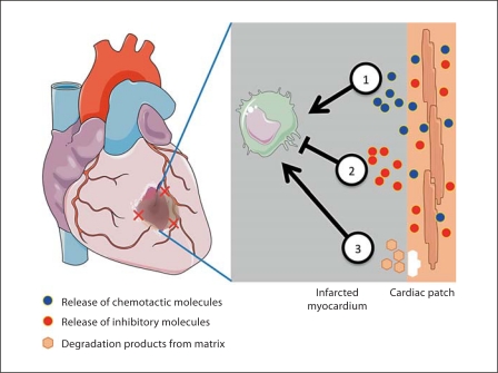
Damaged heart muscle has only a minimal ability for regeneration following myocardial infarction in which cardiomyocytes are lost to ischemia. The most clinically promising approach to regeneration of cardiac muscle currently under investigation is that of injecting cardiogenic repair cells or implanting a preformed tissue-engineered patch. While major advances are being made in the derivation of functional human cardiomyocytes and the development of tissue-engineering modalities for cardiac repair, the host environment into which the repair cells are placed is largely overlooked. Within seconds of myocardial ischemia, hypoxia sets in in the myocardium and the inflammatory response starts, characterized by rapid deployment of circulating cells and the release of paracrine and autocrine signals. Therefore, the inflammatory conditions under which these interactions take place, the design of the scaffold material used, and the maturity of the implanted cells will determine the outcomes of any stem cell-based therapy. We discuss here the interactions between implanted and inflammatory cells of the host, which are critical for the design of effective heart repair therapies.


Figures

Fig. 1
Fig. 1
Examples of two approaches to cardiac tissue engineering. Stem cells are differentiated into cardiac repair cells and seeded onto a scaffold material or mixed with an injectable matrix. The scaffold with the repair cells is placed at the site of MI or used to replace the infarcted tissue. Cells encapsulated within a hydrogel matrix are delivered to the site of injury via direct injection. Created using Servier Medical Art.
Fig. 2
Fig. 2
Dynamic and reciprocal interactions between the infarcted myocardium and the engineered cardiac patch. (1) Infiltrating cells will interact with the delivered cells via direct contact and/or secreted molecules. (2) Degradation products of the scaffold or matrix containing the cells will interact with the host tissue cells and infiltrating cells. (3) The repair cells will also interact with the healthy and necrotic myocardial tissue at the infarct site. Created using Servier Medical Art.
Fig. 3
Fig. 3
Presence of inflammatory cells at the infarct site. Following MI there is a transient presence of different inflammatory cells such as neutrophils, monocytes, and polarized macrophages. Proinflammatory macrophages (M1) appear early during the inflammatory response and their number decreases over time with the eventual increase in anti-inflammatory macrophages (M2). Created using Servier Medical Art.
Fig. 4
Fig. 4
Screening of platforms for optimization of key parameters in cardiac tissue engineering. Screening platforms can use the knowledge gained from in vivo testing in small animals (such as the sequential presence of inflammatory cells) to build more accurate screening platforms that take into account not only the biophysical stimuli present but also the host tissue response. The information gained can be validated in using large animal models that more closely resemble the physiological size and mechanical loading of the human heart. The fundamental goal is to build accurate screening platforms using human cells to predict the potential survival and function of engineered cardiac tissues. Ultimately the objective is to harness the host tissue response and repair the damaged tissue. Created using Servier Medical Art.
Fig. 5
Fig. 5
Harnessing of the inflammatory response to improve the survival and engraftment of engineered cardiac tissues. (1) Chemotactic molecules that recruit supportive inflammatory cells can be encapsulated within the scaffold material and controlled-released into the wound site. (2) Alone or in combination with chemotactic molecules, inhibitory molecules can be encapsulated within the scaffold material to inhibit any cell that does not support the survival and engraftment of the patch. (3) The choice of scaffold material, along with its porosity and composition, and the degradation products that result from the host tissue response mediated breakdown could also provide stimulatory or inhibitory signals. Individually or in combination, the mechanisms shown can be used to harness the inflammatory environment leading to an optimized engineered cardiac patch. Created using Servier Medical Art.

References

    1. Abarbanell A.M., Herrmann J.L., Weil B.R., Wang Y., Tan J., Moberly S.P., Fiege J.W., Meldrum D.R. Animal models of myocardial and vascular injury. J Surg Res. 2010;162:239–249. - PubMed
    1. Ascher N.L., Hoffman R.A., Hanto D.W., Simmons R.L. Cellular basis of allograft rejection. Immunol Rev. 1984;77:217–232. - PubMed
    1. Atluri P., Woo Y.J. Pro-angiogenic cytokines as cardiovascular therapeutics: assessing the potential. BioDrugs. 2008;22:209–222. - PubMed
    1. Auwerx J. The human leukemia cell line, THP-1: a multifacetted model for the study of monocyte-macrophage differentiation. Experientia. 1991;47:22–31. - PubMed
    1. Badylak S.F., Freytes D.O., Gilbert T.W. Extracellular matrix as a biological scaffold material: structure and function. Acta Biomater. 2009;5:1–13. - PubMed

Publication types