

How blood vessel networks are made and measured
- PMID: 21996655
- PMCID: PMC3325601
- DOI: 10.1159/000331398
How blood vessel networks are made and measured
Abstract
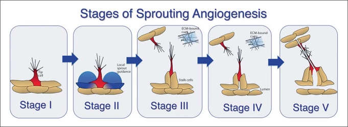
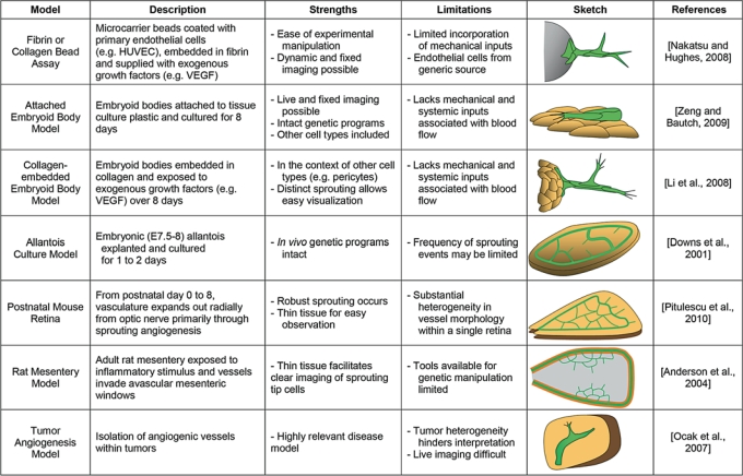
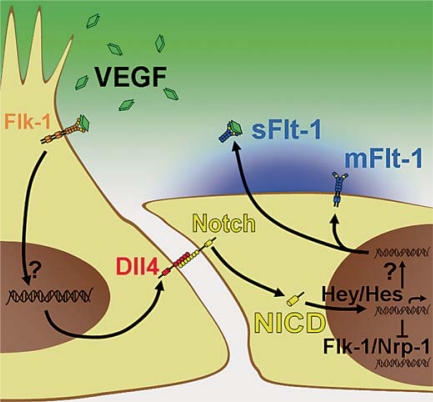
Tissue and organ viability depends on the proper systemic distribution of cells, nutrients, and oxygen through blood vessel networks. These networks arise in part via angiogenic sprouting. Vessel sprouting involves the precise coordination of several endothelial cell processes including cell-cell communication, cell migration, and proliferation. In this review, we discuss zebrafish and mammalian models of blood vessel sprouting and the quantification methods used to assess vessel sprouting and network formation in these models. We also review the mechanisms involved in angiogenic sprouting, and we propose that the process consists of distinct stages. Sprout initiation involves endothelial cell interactions with neighboring cells and the environment to establish a specialized tip cell responsible for leading the emerging sprout. Furthermore, local sprout guidance cues that spatially regulate this outward migration are discussed. We also examine subsequent events, such as sprout fusion and lumenization, that lead to maturation of a nascent sprout into a patent blood vessel.
Copyright © 2011 S. Karger AG, Basel.
Figures
References
-
- Aiello L.P. Targeting intraocular neovascularization and edema – one drop at a time. N Engl J Med. 2008;359:967–969. - PubMed
-
- Almagro S., Durmort C., Chervin-Petinot A., Heyraud S., Dubois M., Lambert O., Maillefaud C., Hewat E., Schaal J.P., Huber P., Gulino-Debrac D. The motor protein myosin-X transports VE-cadherin along filopodia to allow the formation of early endothelial cell-cell contacts. Mol Cell Biol. 2010;30:1703–1717. - PMC - PubMed
-
- Anderson C.R., Ponce A.M., Price R.J. Immunohistochemical identification of an extracellular matrix scaffold that microguides capillary sprouting in vivo. J Histochem Cytochem. 2004;52:1063–1072. - PubMed
-
- Argraves W.S., Larue A.C., Fleming P.A., Drake C.J. VEGF signaling is required for the assembly but not the maintenance of embryonic blood vessels. Dev Dyn. 2002;225:298–304. - PubMed
Publication types
MeSH terms
Grants and funding
LinkOut - more resources
Full Text Sources
