. 2011 Dec;179(6):3020-31.
doi: 10.1016/j.ajpath.2011.08.038. Epub 2011 Oct 11.

Snail1 mediates hypoxia-induced melanoma progression

Affiliations

Snail1 mediates hypoxia-induced melanoma progression

Shujing Liu et al. Am J Pathol. 2011 Dec.

Abstract

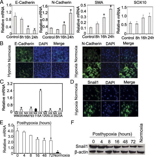
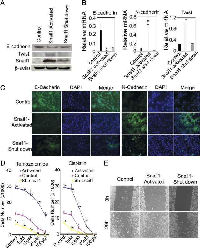
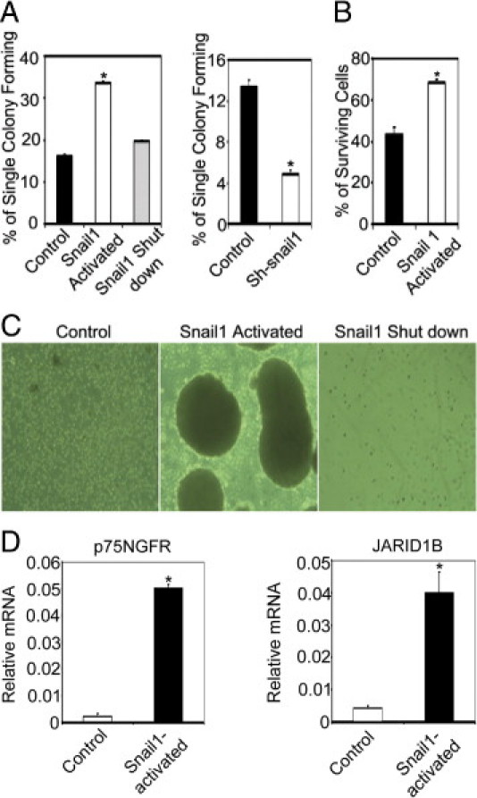
Tumor hypoxia is a known adverse prognostic factor, and the hypoxic dermal microenvironment participates in melanomagenesis. High levels of hypoxia inducible factor (HIF) expression in melanoma cells, particularly HIF-2α, is associated with poor prognosis. The mechanism underlying the effect of hypoxia on melanoma progression, however, is not fully understood. We report evidence that the effects of hypoxia on melanoma cells are mediated through activation of Snail1. Hypoxia increased melanoma cell migration and drug resistance, and these changes were accompanied by increased Snail1 and decreased E-cadherin expression. Snail1 expression was regulated by HIF-2α in melanoma. Snail1 overexpression led to more aggressive tumor phenotypes and features associated with stem-like melanoma cells in vitro and increased metastatic capacity in vivo. In addition, we found that knockdown of endogenous Snail1 reduced melanoma proliferation and migratory capacity. Snail1 knockdown also prevented melanoma metastasis in vivo. In summary, hypoxia up-regulates Snail1 expression and leads to increased metastatic capacity and drug resistance in melanoma cells. Our findings support that the effects of hypoxia on melanoma are mediated through Snail1 gene activation and suggest that Snail1 is a potential therapeutic target for the treatment of melanoma.


Figures

Figure 1
Figure 1
Hypoxia enhances melanoma drug resistance and migration. A: Effect of hypoxia on melanoma cells. WM115A cells were incubated under 1.0% or 21% O2 for 8, 16, 24 hours (n = 3 replicate experiments, *P < 0.05 compared with control). B: Effect of cisplatin and temozolomide on melanoma cells. Normoxia and hypoxia-treated WM115A cells were incubated with 1, 10, or 25 μmol/L of cisplatin and temozolomide (n = 3 replicate experiments; *P < 0.05 compared with control). C: Cell migration assay. Wound healing of normoxia- or hypoxia-treated WM115A cells at 0 and 20 hours after scratch formation. Shown are representative images from three experiments. D: Soft agar assay. Normoxia- or hypoxia-treated WM115A were incubated in soft agar. The clones were counted using a microscope at 100× power. The values (colony number) are expressed as mean ± SD (SD) from three separate measurements (*P < 0.05 compared with normoxia-treated cells).
Figure 2
Figure 2
Hypoxia induces Snail1 up-regulation and E-cadherin down-regulation. A: Quantitative RT-PCR analysis of E-cadherin, N-cadherin, smooth muscle actin (SMA), and SOX10 expression after hypoxia treatment. WM115A cells were cultured under 1.0% or 21% O2 for 8, 16, 24 hours (n = 3 replicate experiments; *P < 0.05 compared with control). B: Expression of E-cadherin or N-cadherin after hypoxia treatment. Immunocytochemical stain of E-cadherin and N-cadherin in WM115A after hypoxia treatment. C: Snail1 gene expression after hypoxia treatment. Snail1 expression in WM35, WM793, WM115A, 1205LU, and WM3523A was assessed by quantitative RT-PCR (n = 3 replicate experiments; *P < 0.01 compared with control). D: Expression of Snail1 by immunocytochemistry (n = 3 replicate experiments). E: Snail1 gene expression in WM115A cells (post-hypoxia 0, 4, 8, 16, 48, and 72 hours) was assessed by quantitative RT-PCR (n = 3 replicate experiments; *P < 0.01 compared with control). F: Expression of Snail1 by Western blot. Cell lysates from WM115A (post-hypoxia 0, 4, 8, 16, 48, and 72 hours) were used for Western blot analyses with anti-Snail1 antibody. β-actin was used as loading controls.
Figure 3
Figure 3
HIF-2α up-regulates Snail1. A: Quantitative RT-PCR assay for HIF-1α and HIF-2α expression in WM115A cells transfected with plasmids containing HIF-1α or HIF-2α (n = 3 replicate experiments; *P < 0.01 compared with control). B: Quantitative RT-PCR assay for Snail1 expression in WM115A cells transfected with plasmids containing HIF-1α or HIF-2α (n = 3 replicate experiments; *P < 0.01 compared with control). C: Western blot analysis for Snail1 expression in WM115A cells transfected with plasmids containing HIF-1α or HIF-2α (n = 3 replicate experiments). D: Quantitative RT-PCR assay for HIF-1α and HIF-2α expression in WM115A cells transfected with plasmids containing Si-HIF-1α or Si-HIF-2α (n = 3 replicate experiments; *P < 0.01 compared with control). E: Quantitative RT-PCR assay for Snail1 expression in WM115A cells transfected with plasmids containing Si-HIF-1α or Si-HIF-2α (n = 3 replicate experiments; *P < 0.01 compared with control). F: Western blot analysis for Snail1 expression in WM115A cells transfected with plasmids containing Si-HIF-1α or Si-HIF-2α (n = 3 replicate experiments).
Figure 4
Figure 4
Effects of Snail1 activation. A: Snail1, E-cadherin, and Twist expression in WM115A cells (n = 3 replicate experiments). Tumor cells were infected with pWZL-Blast-ER-Snail1 and incubated in the medium with absence, presence, or after withdraw of tamoxifen (shutdown of exogenous Snail1 expression). B: Quantitative RT-PCR for E-cadherin, N-cadherin, and Twist expression in WM115A cells with pWZL-Blast-ER-Snail1 (n = 3 replicate experiments; *P < 0.01 compared with control). C: Immunocytochemical stain for E-cadherin and N-cadherin in WM115A cells with pWZL-Blast-ER-Snail1. D: Resistance to temozolamide and cisplatin. The same number of WM115 cells control and WM115 cells with pWZL-Blast-ER-Snail1 or pGIPZ-Snail1 were incubated in medium containing 0, 1, 10, 25, or 100 μmol/L temozolamide or cisplatin for 24 hours (n = 3 replicate experiments; *P < 0.01 compared with control). E: Cell migration assay. Wound healing assay using WM115A with pWZL-Blast-ER-Snail1 at 0 and 20 hours after scratch forming (n = 3 replicate experiments).
Figure 5
Figure 5
Effects of Snail1 knockdown. A: Expression of Snail1. Quantitative RT-PCR for Snail1 expression after Snail1 knockdown using shRNA (sh-snail1) or siRNA (Si-snail1). Experiments were performed under normoxia. (*P < 0.05 compared to control; n = 3 replicate experiments). B: Expression of E-cadherin and N-cadherin after Snail1 knockdown. Immunocytochemical stain for E-cadherin and N-cadherin in Snail1 knockdown and control cells. Experiments were performed under normoxia. C: Western blot analysis for Snail1, E-cadherin, and N-cadherin expression in WM115A cells transfected with Si-Snail1 or Sh-snail1 (n = 3 replicate experiments). Experiments were performed under normoxia. D: Cell cycle analysis. Cells in G2-M and S phases were analyzed by FACS analysis (n = 3 replicate experiments). Experiments were performed under normoxia. E: Cell survival under normoxia and hypoxia. Snail1 knockdown WM115A cells and control cells were cultured under 1% O2 or room O2 for 16 hours (n = 3 replicate experiments; *P < 0.01 compared with control). F: Cell migration assay under normoxia and hypoxia. Wound healing assay using WM115A with Snail1 knockdown (n = 3 replicate experiments).
Figure 6
Figure 6
Snail1 activation induces cancer stem-like cell phenotypes. A: Cell proliferation capacity. Limiting dilution assay was performed and single cells were followed for 14 days using control and WM115A cells with pWZL-Blast-ER-Snail1 (left panel) or control and tumor cells with pGIPZ-Snail1 (right panel). The number of colonies formed was counted and averaged in 3 replicate experiments. (*P < 0.01 comparing to control). B: Hypoxia tolerance assay. Same number of control and WM115A cells with pWZL-Blast-ER-Snail1 were seeded and cultured under 1% O2 for 16 hours (n = 3 replicate experiments). (*P < 0.05 comparing to control). Survival cells were counted. C: Spheroid formation. WM115A cells with pWZL-Blast-ER-Snail1 were cultured in the hESMC4 medium in ultra-low attachment plates. After 2 weeks, photographs were taken of the spheroids at 100× magnification. D: Expression of p75NGFR and JARID1B. Quantitative RT-PCR for p75NGFR and JARID1B expression in sphere-forming cells with pWZL-Blast-ER-Snail1. (*P < 0.05 compared to control; n = 3 replicate experiments).
Figure 7
Figure 7
Snail1 increases melanoma growth and metastasis in vivo. A: Subcutaneous xenografts; 2 × 10⁶ of control, WM115A with pWZL-Blast-ER-Snail1, WM115A with pGIPZ-Snail1 were injected subcutaneously in the flanks of nude mice (n = 10), and these mice were followed for 5 weeks. Representative primary xenografts are shown on the left panel and average tumor weights are shown on the right panel (*P < 0.05 compared with control). B: Internal organ metastasis rate (*P < 0.01 compared with control). C:Snail1, E-cadherin, N-cadherin, and Twist gene expression in xenografts. Quantitative RT-PCR was performed to measure gene expression. D: N-cadherin, Snail1, E-cadherin, and Twist protein expression in the xenografts was assayed using Western blots (representative blot from three experiments).

References

    1. Greenlee R.T., Murray T., Bolden S., Wingo P.A. Cancer statistics, 2000. CA Cancer J Clin. 2000;50:7–33. - PubMed
    1. Wingo P.A., Ries L.A., Giovino G.A., Miller D.S., Rosenberg H.M., Shopland D.R., Thun M.J., Edwards B.K. Annual report to the nation on the status of cancer, 1973–1996, with a special section on lung cancer and tobacco smoking. J Natl Cancer Inst. 1999;91:675–690. - PubMed
    1. Rigel D.S., Carucci J.A. Malignant melanoma: prevention, early detection, and treatment in the 21st century. CA Cancer J Clin. 2000;50:215–236. - PubMed
    2. quiz 237–240

    1. Brizel D.M., Scully S.P., Harrelson J.M., Layfield L.J., Bean J.M., Prosnitz L.R., Dewhirst M.W. Tumor oxygenation predicts for the likelihood of distant metastases in human soft tissue sarcoma. Cancer Res. 1996;56:941–943. - PubMed
    1. Sundfor K., Lyng H., Rofstad E.K. Tumour hypoxia and vascular density as predictors of metastasis in squamous cell carcinoma of the uterine cervix. Br J Cancer. 1998;78:822–827. - PMC - PubMed

Publication types

MeSH terms