. 2012 Jan;5(1):73-81.
doi: 10.1158/1940-6207.CAPR-11-0348. Epub 2011 Oct 13.

Hops (Humulus lupulus) inhibits oxidative estrogen metabolism and estrogen-induced malignant transformation in human mammary epithelial cells (MCF-10A)

Affiliations

Hops (Humulus lupulus) inhibits oxidative estrogen metabolism and estrogen-induced malignant transformation in human mammary epithelial cells (MCF-10A)

L P Hemachandra et al. Cancer Prev Res (Phila). 2012 Jan.

Abstract

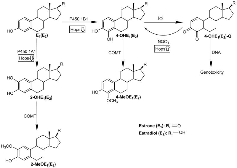
Long-term exposure to estrogens including those in traditional hormone replacement therapy (HRT) increases the risk of developing hormone-dependent cancers. As a result, women are turning to over-the-counter (OTC) botanical dietary supplements, such as black cohosh (Cimicifuga racemosa) and hops (Humulus lupulus), as natural alternatives to HRT. The two major mechanisms which likely contribute to estrogen and/or HRT cancer risk are: the estrogen receptor-mediated hormonal pathway; and the chemical carcinogenesis pathway involving formation of estrogen quinones that damage DNA and proteins, hence initiating and promoting carcinogenesis. Because, OTC botanical HRT alternatives are in widespread use, they may have the potential for chemopreventive effects on estrogen carcinogenic pathways in vivo. Therefore, the effect of OTC botanicals on estrogen-induced malignant transformation of MCF-10A cells was studied. Cytochrome P450 catalyzed hydroxylation of estradiol at the 4-position leads to an o-quinone believed to act as the proximal carcinogen. Liquid chromatography/tandem mass spectrometry analysis of estradiol metabolites showed that 4-hydroxylation was inhibited by hops, whereas black cohosh was without effect. Estrogen-induced expression of CYP450 1B1 and CYP450 1A1 was attenuated by the hops extract. Two phenolic constituents of hops (xanthohumol, XH; 8-prenylnaringenin, 8-PN) were tested: 8-PN was a potent inhibitor, whereas XH had no effect. Finally, estrogen-induced malignant transformation of MCF-10A cells was observed to be significantly inhibited by hops (5 μg/mL) and 8-PN (50 nmol/L). These data suggest that hops extracts possess cancer chemopreventive activity through attenuation of estrogen metabolism mediated by 8-PN.


Figures

Fig. 1
Fig. 1
Scheme of potential mechanisms by which hops can modulate estrogen metabolism in breast epithelial cells. Hops inhibits E2 induced upregulation of CYP450 1A1 and 1B1 resulting reduced formation of catechol estrogens leading to less quinone production; hence, DNA damage and malignant transformation is decreased. Since catechol O-methyltransferase (COMT) converts 2-hydroxyestrone and 4-hydroxyestrone into the corresponding 2- and 4-methoxy metabolites, these served as indices of catechol estrogen formation.
Fig. 2
Fig. 2
Effect of black cohosh and hops on oxidative estrogen metabolism in MCF-10A cells. Positive ion-electrospray selective reaction monitoring (SRM) chromatograms of 2-MeOE1 and 4-MeOE1. (A) Black cohosh had no significant effect on 2-MeOE1 and 4-MeOE1 formation while (B) hops significantly inhibited the formation of 2-MeOE1 and 4-MeOE1. MCF-10 A cells were treated with and without black cohosh (20 μg/mL) or hops (5 μg/mL) in the presence of E2 (1 μM) for 6 days. Cell media was collected and the metabolites were extracted in to dichloromethane followed by the derivatization using dansyl chloride. Samples were analyzed by LC-MS/MS in multiple reaction monitoring (MRM) mode as described in Materials and Methods. Overlaid SRM chromatograms represent the formation of 2-MeOE1 and 4-MeOE1 in E2 treated (line) and E2 and (A) black cohosh (dashed line), or (B) hops (dotted line) treated samples.
Fig. 3
Fig. 3
Dose dependent effect of hops on the formation of 2-MeOE1 and 4-MeOE1. MCF-10A cells were treated with different concentrations of hops in the presence of E2 (1 μM) for 6 days and cell media was collected and analyzed for the estrogen metabolites using LC-MS/MS. Each value represents the average ± SD of three experiments performed independently in duplicate.
Fig. 4
Fig. 4
Hops inhibits E2 induced (A) CYP450 1B1 and (B) 1A1 expression. (C) COMT expression is not significantly affected by either E2 or hops. Cells were collected at different time points (1, 3, 6 days) after the treatment and protein was extracted and analyzed using immunoblotting. Anti CYP450 1B1, anti CYP450 1A1 and anti COMT antibodies were used in 1:200, 1:1000 and 1:1000 dilutions, respectively. These blots are representatives of three experiments done independently. The intensity of the bands was normalized to β-actin as outlined in Material and Methods and represented as relative protein expression. Each lane contains 30 μg of total protein as determined by BCA assay.
Fig. 4
Fig. 4
Hops inhibits E2 induced (A) CYP450 1B1 and (B) 1A1 expression. (C) COMT expression is not significantly affected by either E2 or hops. Cells were collected at different time points (1, 3, 6 days) after the treatment and protein was extracted and analyzed using immunoblotting. Anti CYP450 1B1, anti CYP450 1A1 and anti COMT antibodies were used in 1:200, 1:1000 and 1:1000 dilutions, respectively. These blots are representatives of three experiments done independently. The intensity of the bands was normalized to β-actin as outlined in Material and Methods and represented as relative protein expression. Each lane contains 30 μg of total protein as determined by BCA assay.
Fig. 5
Fig. 5
Effect of XH and 8-PN on estrogen metabolism in MCF-10A cells. 2-MeOE1 (open circles) and 4-MeOE1 (closed circles) formation was plotted against different concentrations of XH and 8-PN. MCF-10A cells were treated with E2 (1 μM) in the presence and absence of different concentrations of either XH or 8-PN for 6 days. Cell media was analyzed for estrogen metabolites using LC-MS/MS. There was a significant inhibition (p< 0.0005) of both 2-MeOE1 and 4-MeOE1 formation in the presence of nanomolar concentrations of 8-PN. Each value represents an average ± SD of three experiments performed independently in duplicate.
Fig. 6
Fig. 6
Effect of hops and 8-PN on E2 induced malignant transformation in MCF-10A cells. MCF-10A cells were treated with E2 (1 μM) in the presence and absence of hops (5 μg/mL) or 8-PN (50 nM). Treatments were continued for 3 weeks and cells were passaged once a week. At the end of 3 weeks, cells were plated and maintained on soft agar for 3 weeks and formation of colonies were observed and counted using an inverted microscope. Hops (p <0.005) and 8-PN (p<0.0001) significantly inhibited E2 induced malignant transformation in MCF-10A cells. DMSO treated (0.01%) samples were used as a negative control. Each point represents an average of triplicate ± SD.

Similar articles

Cited by

References

    1. Russo J, Hasan Lareef M, Balogh G, Guo S, Russo IH. Estrogen and its metabolites are carcinogenic agents in human breast epithelial cells. J Steroid Biochem Mol Biol. 2003;87:1–25. - PubMed
    1. Yaghjyan L, Colditz G. Cancer Causes and Control. Springer; Netherlands: 2011. Estrogens in the breast tissue: a systematic review; pp. 529–40. - PMC - PubMed
    1. Cavalieri EL, Rogan EG. Depurinating estrogen-DNA adducts in the etiology and prevention of breast and other human cancers. Future Oncol. 2010;6:75–91. - PMC - PubMed
    1. Bolton JL. Mechanisms of Estrogen Carcinogenesis: Modulation by Botanical Natural Products. In: Penning TM, editor. Chemical Carcinogenesis. Humana Press; 2011. pp. 75–93.
    1. Bolton JL, Thatcher GRJ. Potential mechanisms of estrogen quinone carcinogenesis. Chem Res Toxicol. 2008;21:93–101. - PMC - PubMed

Publication types