Review
. 2011 Oct 28;12(11):1118-26.
doi: 10.1038/embor.2011.193.

The emerging functions of septins in metazoans

Affiliations
Review

The emerging functions of septins in metazoans

Juha Saarikangas et al. EMBO Rep. .

Abstract

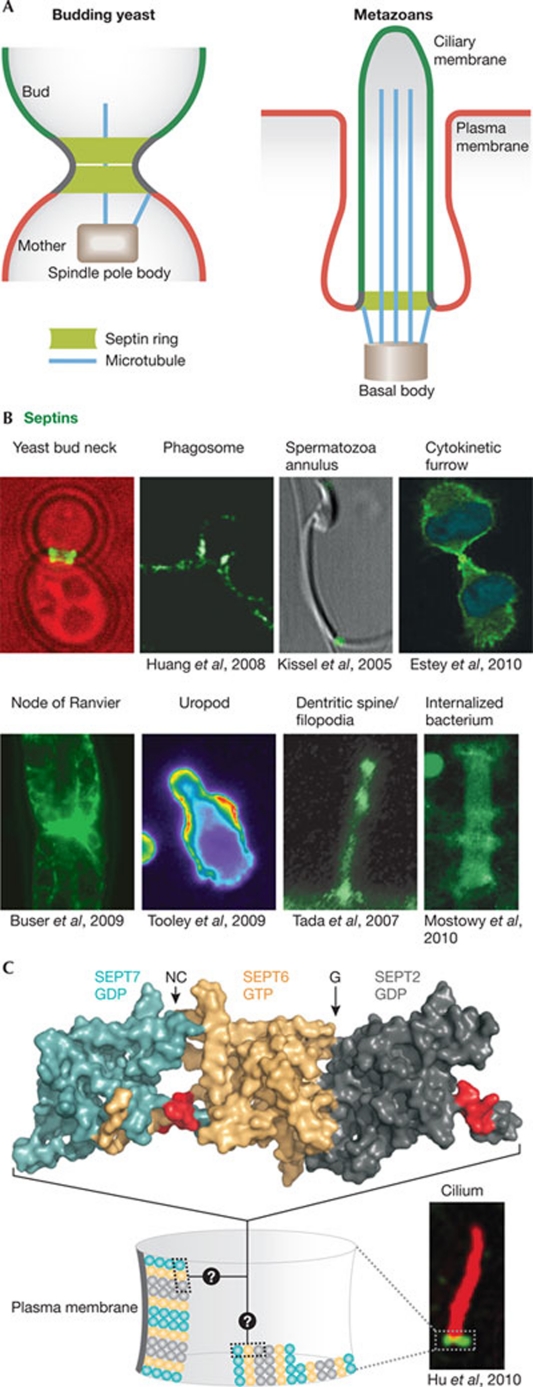
Septins form a subfamily of highly related GTP-binding proteins conserved from eukaryotic protists to mammals. In most cases, septins function in close association with cell membranes and the actin and microtubule cytoskeleton to regulate a wide variety of key cellular processes. Further underscoring their importance, septin abnormalities are associated with several human diseases. Remarkably, septins have the ability to polymerize into assemblies of different sizes in vitro and in vivo. In cells, these structures act in the formation of diffusion barriers and scaffolds that maintain subcellular polarity. Here, we focus on the emerging roles of vertebrate septins in ciliogenesis, neurogenesis, tumorigenesis and host-pathogen interactions, and discuss whether unifying themes underlie the molecular function of septins in health and disease.


Conflict of interest statement

The authors declare that they have no conflict of interest.

Figures

Figure 1
Figure 1
The septin ring paradigm. (A) Septins assemble at the neck of budding yeast cells in a ring-like collar. This functions as a diffusion barrier to control the exchange of molecules between the mother and the bud, and as a scaffold to anchor other proteins, such as astral microtubules emanating from the spindle pole body (centriole equivalent in yeast). Cilia contain a structurally and functionally analogous septin structure at their base. (B) In metazoans, membrane-associated septin assemblies are found at the base of the indicated cellular structures and around intracellular pathogens (Shigella flexneri). (C) Structure of the SEPT2 (turquoise)–SEPT6 (gold)–SEPT7 (dark grey) complex (Protein Data Bank: 2QAG; Sirajuddin et al, 2007). The proposed membrane-binding interface (polybasic region; Zhang et al, 1999) is shown in red and the arrows denote the NC- and G-oligomerization interfaces. When assembled into rings, these oligomers probably form filaments, but the orientation of filaments in metazoan septin rings is unknown. Images in panels B and C are reproduced with permission from the relevant publishers. SEPT, septin.
Figure 2
Figure 2
Possible roles for septins in cilia and neuronal synapses. (A) Septins might contribute to ciliogenesis in different ways. (1) The septin ring acts as a lateral diffusion barrier at the base of the cilium and controls the transport of membrane proteins. (2) The septin ring could provide the attachment site for the transition fibres (orange), directly or through adaptor proteins such as APC. (3) The septin ring might function as a scaffold to recruit and/or organize the function of the exocyst, PCP proteins and the BBSome. (4) Septins are also present at the tip and shaft of the cilium and could influence microtubule dynamics and the IFT. (5) Septins could also function in vesicle transport to the basal body. The red colour at the base of the cilium indicates the ciliary necklace region, which displays a high degree of positive membrane curvature, possibly recognized by amphipathic helices (such as in the BBSome subunit Arl6). (B) (1) Septins localize to the base of the dendritic spines, where they might act as lateral diffusion barriers to restrict exchange between the dendritic shaft and the spine. Septins could also influence spine morphogenesis by regulating (2) actomyosin structures or (3) microtubules. Moreover, (4) septins are enriched in the PSD and (5) hinder synaptic vesicle release at the presynaptic compartment by acting as membrane-apposed barriers. APC, adenomatous polyposis coli; BBSome, Bardet–Biedel syndrome proteins; IFT, intraflagellar transport; PCP, planar cell polarity; PSD, postsynaptic density.
Figure 3
Figure 3
Interplay between septins and the actin and microtubule cytoskeletons. (A) Septins localize to microtubules and might influence them in different ways: (1) guide microtubule growth and prevent their disassembly; (2) influence microtubule acetylation; (3) bind to polyglutamated microtubules; (4) act as scaffolds to recruit other proteins to microtubules; (5) regulate the motility of microtubule-driven motors such as CENP-E and influence vesicle transport along microtubules; and (6) interact with MAPs to control the dynamics and identity of microtubules. (B) In mammalian cells, some septins localize to actomyosin structures, including stress fibres, contractile rings and around actomyosin-covered intracellular bacteria. Septins are recruited to stress fibres by binding to NM2, and this interaction might be enhanced by different factors such as TNFα. Septins recruit ROCK and CRIK to promote MLC phosphorylation and actomyosin contractility, and can possibly recruit formins to promote actomyosin assembly. Images in panels A and B are reproduced with permission from the relevant publishers. CENP-E, centromere-associated protein E; CRIK, citron kinase; MAP, microtubule-associated protein; MLC, myosin light chain kinase; NM2, type II non-muscle myosin; TNFα, tumour necrosis factor α; ROCK, Rho kinase.

Similar articles

  • Septins: the fourth component of the cytoskeleton.
    Mostowy S, Cossart P. Mostowy S, et al. Nat Rev Mol Cell Biol. 2012 Feb 8;13(3):183-94. doi: 10.1038/nrm3284. Nat Rev Mol Cell Biol. 2012. PMID: 22314400 Review.
  • Cell and molecular biology of septins.
    Fung KY, Dai L, Trimble WS. Fung KY, et al. Int Rev Cell Mol Biol. 2014;310:289-339. doi: 10.1016/B978-0-12-800180-6.00007-4. Int Rev Cell Mol Biol. 2014. PMID: 24725429 Review.
  • [Functional Characterization of Septin Complexes].
    Akhmetova KA, Chesnokov IN, Fedorova SA. Akhmetova KA, et al. Mol Biol (Mosk). 2018 Mar-Apr;52(2):155-171. doi: 10.7868/S0026898418020015. Mol Biol (Mosk). 2018. PMID: 29695686 Free PMC article. Review.
  • Uncovering the Roles of Septins in Cilia.
    Palander O, El-Zeiry M, Trimble WS. Palander O, et al. Front Cell Dev Biol. 2017 Apr 6;5:36. doi: 10.3389/fcell.2017.00036. eCollection 2017. Front Cell Dev Biol. 2017. PMID: 28428954 Free PMC article. Review.
  • Unearthing the role of septins in viral infections.
    Khairat JE, Hatta MNA, Abdullah N, Azman AS, Calvin SYM, Syed Hassan S. Khairat JE, et al. Biosci Rep. 2024 Mar 29;44(3):BSR20231827. doi: 10.1042/BSR20231827. Biosci Rep. 2024. PMID: 38372298 Free PMC article. Review.

Cited by

References

    1. Amin ND, Zheng YL, Kesavapany S, Kanungo J, Guszczynski T, Sihag RK, Rudrabhatla P, Albers W, Grant P, Pant HC (2008) Cyclin-dependent kinase 5 phosphorylation of human septin SEPT5 (hCDCrel-1) modulates exocytosis. J Neurosci 28: 3631–3643 - PMC - PubMed
    1. Ashby MC, Maier SR, Nishimune A, Henley JM (2006) Lateral diffusion drives constitutive exchange of AMPA receptors at dendritic spines and is regulated by spine morphology. J Neurosci 26: 7046–7055 - PMC - PubMed
    1. Barral Y, Mermall V, Mooseker MS, Syder M (2000) Compartmentalization of the cell cortex by septins is required for the maintenance of cell polarity in yeast. Mol Cell 5: 841–851 - PubMed
    1. Beites CL, Xie H, Bowser R, Trimble WS (1999) The septin CDCrel-1 binds syntaxin and inhibits exocytosis. Nat Neurosci 2: 434–439 - PubMed
    1. Beites CL, Campbell KA, Trimble WS (2005) The septin Sept5/CDCrel-1 competes with α-SNAP for binding to the SNARE complex. Biochem J 385: 347–353 - PMC - PubMed

Publication types