. 2012 Jan;60(1):29-38.
doi: 10.2310/JIM.0b013e318237eb55.

Protein kinase C-beta inhibition induces apoptosis and inhibits cell cycle progression in acquired immunodeficiency syndrome-related non-hodgkin lymphoma cells

Affiliations

Protein kinase C-beta inhibition induces apoptosis and inhibits cell cycle progression in acquired immunodeficiency syndrome-related non-hodgkin lymphoma cells

Nakhle S Saba et al. J Investig Med. 2012 Jan.

Abstract

Introduction: Acquired immunodeficiency syndrome (AIDS)-related non-Hodgkin lymphoma (NHL) constitutes an aggressive variety of lymphomas characterized by increased extranodal involvement, relapse rate, and resistance to chemotherapy. Protein kinase C-beta (PKCβ) targeting showed promising results in preclinical and clinical studies involving a wide variety of cancers, but studies describing the role of PKCβ in AIDS-NHL are primitive if not lacking.

Methods: In the present study, 3 AIDS-NHL cell lines were examined: 2F7 (AIDS-Burkitt lymphoma), BCBL-1 (AIDS-primary effusion lymphoma), and UMCL01-101 (AIDS-diffuse large B-cell lymphoma).

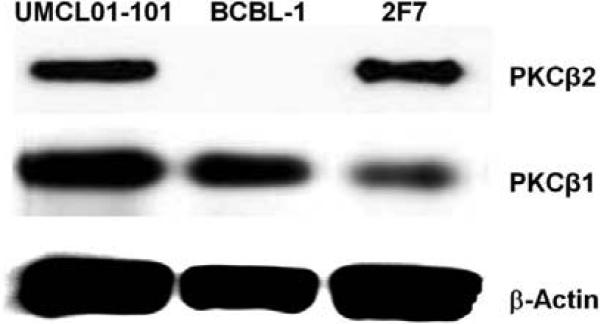
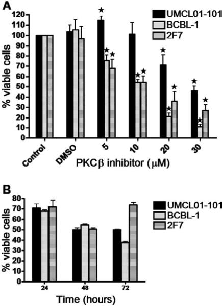
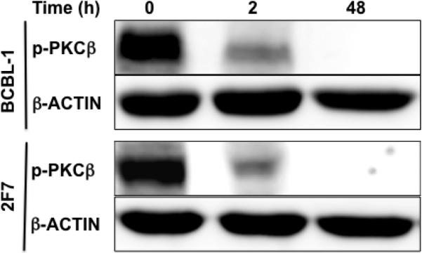
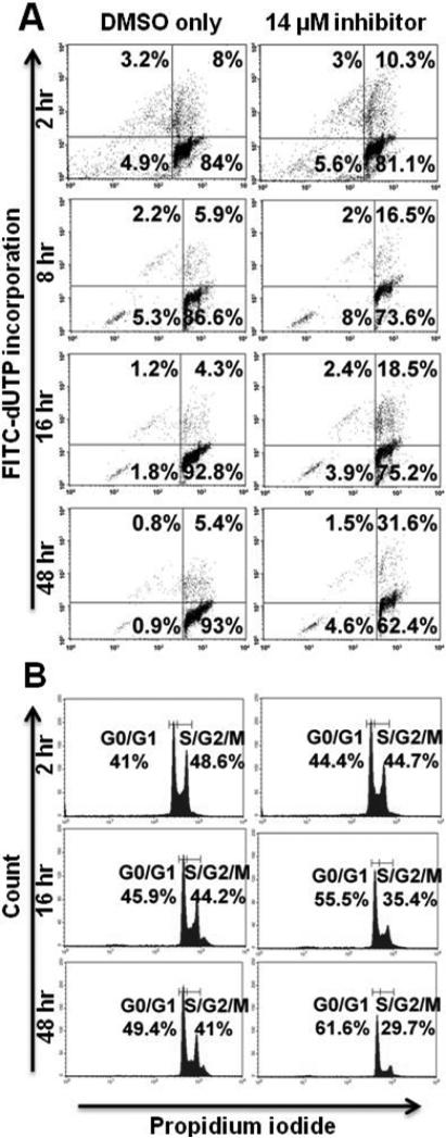
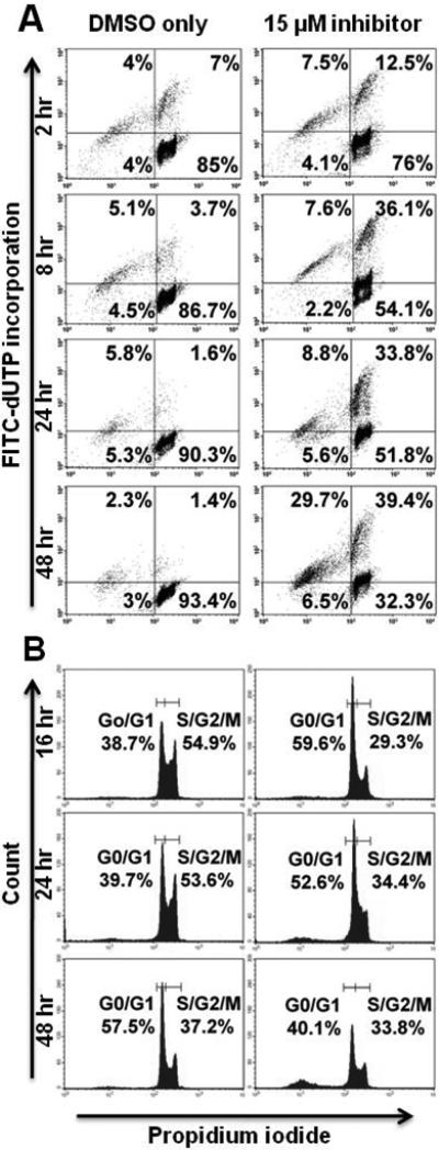
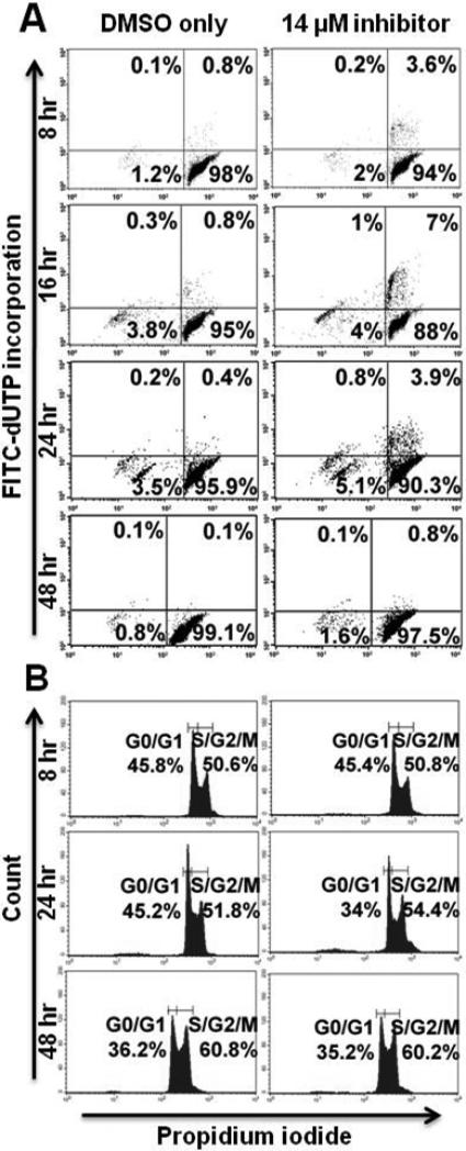
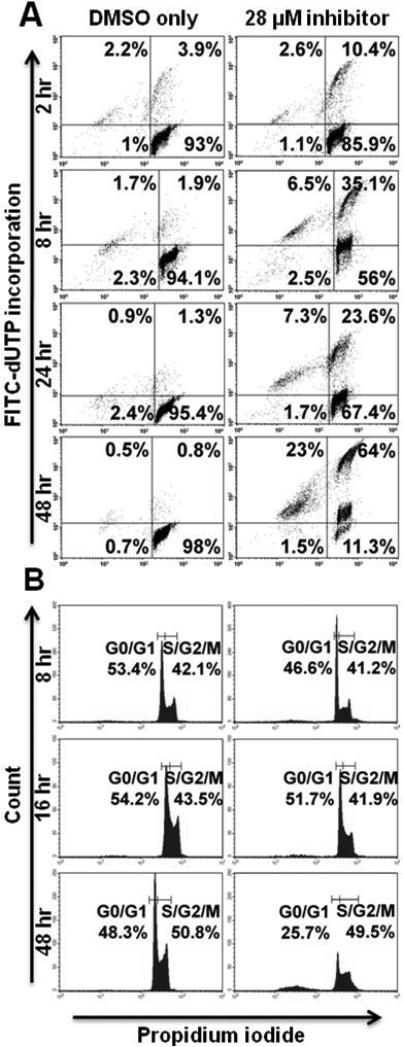
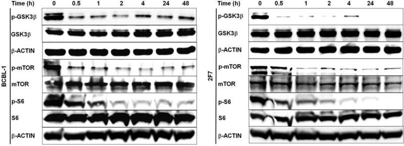
Results: Immunoblot analysis demonstrated expression of PKCβ1 and PKCβ2 in 2F7 and UMCL01-101 cells, and PKCβ1 alone in BCBL-1 cells. The viability of 2F7 and BCBL-1 cells decreased significantly in the presence of PKCβ-selective inhibitor at half-maximal inhibitory concentration of 14 and 15 μmol/L, respectively, as measured by tetrazolium dye reduction assay. In contrast, UMCL01-101 cells were relatively resistant. As determined using flow cytometric deoxynucleotidyl transferase dUTP nick-end labeling assay with propidium iodide staining, the responsiveness of sensitive cells was associated with apoptotic induction and cell cycle inhibition. Protein kinase C-beta-selective inhibition was observed not to affect AKT phosphorylation but to induce a rapid and sustained reduction in the phosphorylation of glycogen synthase kinase-3 beta, ribosomal protein S6, and mammalian target of rapamycin in sensitive cell lines.

Conclusions: The results indicate that PKCβ plays an important role in AIDS-related NHL survival and suggest that PKCβ targeting should be considered in a broader spectrum of NHL. The observations in BCBL-1 were unexpected in the absence of PKCβ2 expression and implicate PKCβ1 as a regulator in those cells.


Figures

Figure 1
Figure 1. Immunoblot analysis of PKCβ1 and PKCβ2 expression in UMCL01-101, BCBL-1 and 2F7 cell lines
Total cellular protein (100 μg) from each cell line was examined first using monoclonal antibody for PKCβ2. The signal was then stripped, followed by analysis with monoclonal antibody for PKCβ1. The expression of β-Actin was examined as a loading control.
Figure 2
Figure 2. Reduction in viability of AIDS-NHL cell lines following PKCβ-selective inhibition
(A) UMCL01-101, BCBL-1 and 2F7 cells were incubated with increasing concentrations of PKCβ-selective inhibitor (0 – 30 μM) for 48 hours, or were treated with the concentration of DMSO vehicle corresponding to the amount present in each dilution of inhibitor stock. Cell viability was quantified using a tetrazolium dye reduction assay. Data are expressed as percentage of the corresponding DMSO-treated control. Shown are data from untreated cells (Control), from cells treated with DMSO concentration equivalent to 30 μM inhibitor (DMSO), and from treatment with inhibitor at 5 – 30 μM. Error bars represent standard deviation. The asterisk (*) indicates statistically significant difference (p < 0.05) as compared to the respective DMSO-treated control. (B) UMCL01-101, BCBL-1 and 2F7 cells were incubated with correspondent IC50 of PKCβ-selective inhibitor for 24, 48 or 72 hours. Cell viability was quantified using a tetrazolium dye reduction assay, and data are expressed as percentage of control cells treated with DMSO only.
Figure 3
Figure 3. Immunoblot analysis of phospho-PKCβ expression in BCBL-1 and 2F7 cell lines
BCBL-1 and 2F7 cells were treated with PKCβ-selective inhibitor at the IC50 (15 μM or 14 μM, respectively) for 2 – 48 hours. Total cellular protein (50 μg) collected at regular intervals was examined using affinity-purified polyclonal antibody for phospho-PKCβ1&2(threonine500). The expression of β-Actin was examined as a loading control.
Figure 4
Figure 4. PKCβ-selective inhibition induces apoptosis and inhibits cell cycle progression in 2F7 cells
2F7 cells were treated with PKCβ-selective inhibitor at the IC50 (14 μM), or with the correspondent concentration of DMSO, for 2 – 48 hours. (A) Cells were examined at regular intervals for apoptosis using a flow cytometric TUNEL assay in which apoptotic cells are demonstrated in the right and left upper quadrants. (B) Cell cycle analysis was performed at regular intervals using propidium iodide staining. Representative histograms are shown.
Figure 5
Figure 5. PKCβ-selective inhibition induces apoptosis and transiently inhibits cell cycle progression in BCBL-1 cells
BCBL-1 cells were treated with PKCβ-selective inhibitor at the IC50 (15 μM), or with the correspondent concentration of DMSO, for 2 – 48 hours. Analyses of apoptosis (A) and cell cycle progression (B) were performed as described for Figure 4.
Figure 6
Figure 6. Relative resistance of UMCL01-101 cells to apoptotic induction or cell cycle inhibition by PKCβ-selective inhibition
UMCL01-101 cells were treated with PKCβ-selective inhibitor at a concentration comparable to the IC50 for 2F7 or BCBL-1 cells (14 μM), or with the correspondent concentration of DMSO, for 2 – 48 hours. Analyses of apoptosis (A) and cell cycle progression (B) were performed as described for Figure 4.
Figure 7
Figure 7. PKCβ-selective inhibition at high concentration induces apoptosis but does not inhibit cell cycle progression in UMCL01-101 cells
UMCL01-101 cells were treated with PKCβ-selective inhibitor at the IC50 (28 μM), or with the correspondent concentration of DMSO, for 2 – 48 hours. Analyses of apoptosis (A) and cell cycle progression (B) were performed as described for Figure 4.
Figure 8
Figure 8. PKCβ-selective inhibition suppresses GSK3β, mTOR and S6 phosphorylation
BCBL-1 and 2F7 cells were treated with correspondent IC50 of the PKCβ-selective inhibitor, or with DMSO only, for the time periods shown. Immunoblot analyses were performed on total protein (50 μg) from cells collected at each time interval, using antibodies specific for p-GSK3β(serine9), p-mTOR(serine2448), p-S6(serine240/244), and total GSK3β, m-TOR, and S6. The expression of β-Actin was examined as a loading control.

Similar articles

Cited by

References

    1. Faivre S, Djelloul S, Raymond E. New paradigms in anticancer therapy: targeting multiple signaling pathways with kinase inhibitors. Semin Oncol. 2006;33:407–420. - PubMed
    1. Leonard JP, Martin P, Barrientos J, et al. Targeted treatment and new agents in diffuse large B-cell lymphoma. Semin Hematol. 2008;45:S11–16. - PubMed
    1. Serova M, Ghoul A, Benhadji KA, et al. Preclinical and clinical development of novel agents that target the protein kinase C family. Semin Oncol. 2006;33:466–478. - PubMed
    1. Wood C, Harrington W., Jr AIDS and associated malignancies. Cell Res. 2005;15:947–952. - PubMed
    1. Aoki Y, Tosato G. Neoplastic conditions in the context of HIV-1 infection. Curr HIV Res. 2004;2:343–349. - PubMed

Publication types

MeSH terms