. 2011 Nov;7(11):1359-70.
doi: 10.4161/auto.7.11.17660. Epub 2011 Nov 1.

Autophagy stimulation by rapamycin suppresses lung inflammation and infection by Burkholderia cenocepacia in a model of cystic fibrosis

Affiliations

Autophagy stimulation by rapamycin suppresses lung inflammation and infection by Burkholderia cenocepacia in a model of cystic fibrosis

Basant A Abdulrahman et al. Autophagy. 2011 Nov.

Abstract

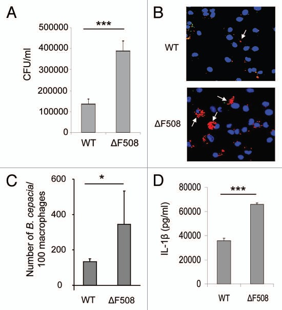
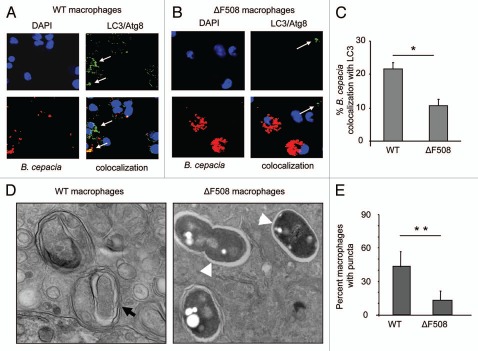
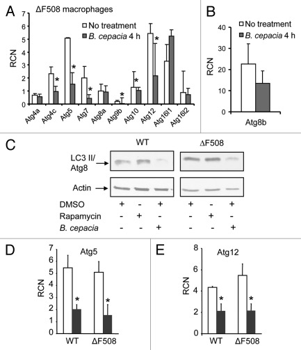
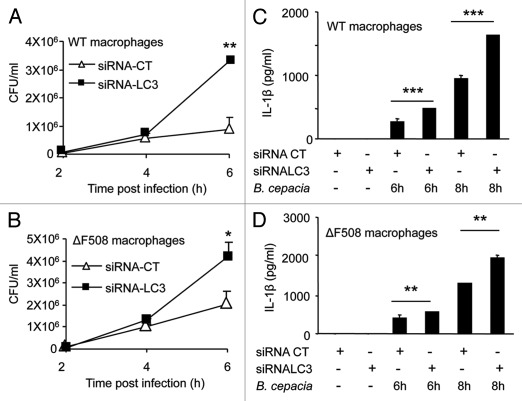
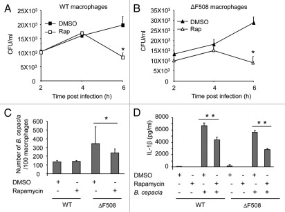
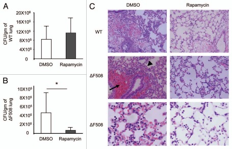
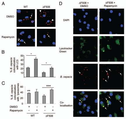
Cystic fibrosis (CF) is the most common inherited lethal disease of Caucasians which results in multi organ dysfunction. However, 85% of the deaths are due to pulmonary infections. Infection by Burkholderia cenocepacia (B. cepacia) is a particularly lethal threat to CF patients because it causes severe and persistent lung inflammation and is resistant to nearly all available antibiotics. In CFTR ΔF508 mouse macrophages, B. cepacia persists in vacuoles that do not fuse with the lysosomes and mediates increased production of IL-1β. It is believed that intracellular bacterial survival contributes to the persistence of the bacterium. Here we show for the first time that in wild-type macrophages but not in ΔF508 macrophages, many B. cepacia reside in autophagosomes that fuse with lysosomes at later stages of infection. Accordingly, association and intracellular survival of B. cepacia are higher in CFTR-ΔF508 (ΔF508) macrophages than in WT macrophages. An autophagosome is a compartment that engulfs non-functional organelles and parts of the cytoplasm then delivers them to the lysosome for degradation to produce nutrients during periods of starvation or stress. Furthermore, we show that B. cepacia downregulates autophagy genes in WT and ΔF508 macrophages. However, autophagy dysfunction is more pronounced in ΔF508 macrophages since they already have compromised autophagy activity. We demonstrate that the autophagy-stimulating agent, rapamycin markedly decreases B. cepacia infection in vitro by enhancing the clearance of B. cepacia via induced autophagy. In vivo, Rapamycin decreases bacterial burden in the lungs of CF mice and drastically reduces signs of lung inflammation. Together, our studies reveal that if efficiently activated, autophagy can control B. cepacia infection and ameliorate the associated inflammation. Therefore, autophagy is a novel target for new drug development for CF patients to control B. cepacia infection and accompanying inflammation.


Figures

Figure 1
Figure 1
Macrophages harboring ΔF508 mutation show more B. cepacia recovery and enhanced cytokine production from macrophages. (A) Wild-type (Wt) or CFTRΔF508 (ΔF508) murine bone marrow-derived macrophages (BMDM) were infected with B. cepacia and colony-forming units (CFUs) were enumerated at 24 h post-infection. (B) confocal microscopy of WT or ΔF508 BMDM after 2 h of infection with B. cepacia. Nuclei are stained blue with DAPI and B. cepacia expresses red florescent protein. White arrows indicate the sites of B. cepacia. (C) scoring of the number of B. cepacia associated with 100 BMDMs in confocal images. (D) 24 h post-infection, supernatants from BMDM from WT or ΔF508 mice were analyzed for IL-1β. Data are representative of 3 independent experiments and presented as the means ± SD. Asterisks in (C and E) indicate significant differences (*p < 0.05 and ***p < 0.0001).
Figure 2
Figure 2
B. cepacia resides in an autophagosome-like compartment in macrophages. (A and B) confocal microscopy for wild-type (WT) murine BMDM (A) and BMDM harboring ΔF508 mutation (ΔF508) (B) representing the colocalization of B. cepacia with LC3/Atg8I after 2 h infection. Nuclei are stained with DAPI, B. cepacia expresses red florescent protein and LC3/Atg8 is stained green with specific antibodies. White arrows indicate the presence of LC3/Atg8. (C) the percentage of colocalization of B. cepacia with LC3/Atg8 was scored. (D) transmission electron microscopy of murine BMDM expressing WT or mutated ΔF508 CFtr protein after 2 h infection with B. cepacia. Black arrow indicates a mutilamellar autophagosome-like vacuole. White arrow heads indicate vacuoles containing intact bacteria within single-membrane vacuoles. (E) the scores for the percentage of puncta in 100 WT and ΔF508 macrophages after 4 h infection with B. cepacia. Data are representative of 3 independent experiments and presented as the means ± SD. Asterisks in (C and E) indicate significant differences (*p < 0.05; **p < 0.01).
Figure 3
Figure 3
B. cepacia downregulates the expression of autophagy genes in murine macrophages. (A and B) Bone marrow-derived macrophages (BMDMs) harboring the ΔF508 mutation were either uninfected (No treatment) or infected with B. cepacia for 4 h and then the cells were lysed in trizol. RNA was extracted and SA Biosciences autophagy array analysis was performed for several autophagy genes (A) and for LC3/Atg8 (B). (C) Immunoblotting for WT and ΔF508 murine macrophages showing the expression level of LC3/Atg8 before and after 4 h infection with B. cepacia in the presence of rapamycin or DMSO. (D and E) q-PCR results for autophagy regulating genes 5 and 12 (Atg5, Atg12) in WT and ΔF508 BMDMs non infected (white bars) or infected (gray bars) with B. cepacia for 4 h. Data in (A, D and E) are expressed as relative copy numbers (RCN) and presented as means of 3 independent experiments ± SD. Asterisks indicate significant differences from the uninfected cells at the indicated time point (*p < 0.05).
Figure 4
Figure 4
Depletion of LC3 with specific siRNA increases B. cepacia multiplication and Interleukin-1β (IL-1β) secretion in macrophages. (A and B) Bone-marrow derived macrophages (BMDMs) from WT and ΔF508 mutant mice were nucleofected with siRNA against LC3/Atg8 (siRNA-LC3) or control siRNA (siRNA-CT) for 48 h and then infected with B. cepacia for 2, 4 and 6 h. colony forming units (CFUs) were enumerated. (C and D) Both types of macrophages were then infected for 6 and 8 h and supernatants were analyzed for IL-1β production. Data are representative of three different experiments and presented as the means ± SD. Asterisks indicate significant differences from the (siRNA-CT) at the respective time points (*p < 0.05; **p < 0.01; ***p< 0.001).
Figure 5
Figure 5
Rapamycin treatment decreases the recovery of B. cepacia and Interleukin-1β (IL-1β) production from infected macrophages. (A and B) Bone-marrow derived macrophages (BMDMs) from WT mice (A) and those harboring the ΔF508 mutation (B) were infected with B. cepacia for 2, 4 and 6 h in the presence of rapamycin or DMSO. Colony-forming units (CFUs) were enumerated. (C) WT and ΔF508 were infected with B. cepacia for 2 h in the presence of rapamycin or DMSO and then the number of bacteria per 100 macrophages was scored. (D) WT and ΔF508 were infected with B. cepacia for 24 h in the presence of rapamycin or DMSO and the supernatants were analyzed for IL-1β. Data are representative of 3 independent experiments and presented as means ± SD. Asterisks indicate significant differences from the DMSO-treated cells (*p < 0.05; **p < 0.01).
Figure 6
Figure 6
Treatment with rapamycin markedly decreases the recovery of B. cepacia from infected lungs of mice harboring ΔF508 mutation in vivo. Wild-type (WT) (A) and mice harboring the ΔF508 mutation (ΔF508) (B) were pretreated with 2 doses of rapamycin (4 mg/kg) or with DMSO at a 24 h interval by intra-peritoneal injections. then, mice were infected intra-tracheally with B. cepacia followed by a dose of rapamycin or DMSO. colony-forming units (CFUs) recovered from homogenized lungs were enumerated and expressed as CFU per gram of lung tissue (A and B). (C) h&e staining of lung sections from WT (upper parts X40) or ΔF508 mice (middle parts X40) treated as in (A and B). Lower part shows higher magnification (X100) of infected ΔF508 lung sections. Data in (A and B) are represented as the means of data obtained from 3 mice ± SD. Asterisks indicate significant differences from the DMSO treated mice (*p < 0.05).
Figure 7
Figure 7
Treatment with rapamycin increases B. cepacia colocalization with LC3/Atg8 and with lysosomes. Bone marrow derived macrophages (BMDM) from wild-type (WT) and BMDM harboring ΔF508 mutation (ΔF508) were treated with rapamycin or DMSO for 1 h before infection, and then macrophages were infected with B. cepacia for 2 h. (A) confocal microscopy demonstrates nuclei stained with DAPI, LC3 stained green and B. cepacia expressing monomeric red florescent protein (mRFP). White arrows indicate the sites of colocalization with of B. cepacia with LC3 with or without rapamycin. White arrows indicate colocalization, white arrow heads indicate B. cepacia. (B) scoring of the percentage of colocalization of B. cepacia with LC3 with or without rapamycin treatment. (C) scoring of the percentage of colocalization of B. cepacia with Lystracker Green with or without rapamycin. (D) confocal microscopy showing nuclei stained with DAPI, lysosomes with Lysotracker Green and mRFP-expressing B. cepacia. the site of colocalization of B. cepacia with Lysotracker Green in ΔF508 macrophages upon rapamycin (ΔF508 + rapamycin) or DMSO (ΔF508 + DMSO) are indicated with white arrows. Data are representative of 3 independent experiments and presented as the means ± SD. Asterisks in (B and c) indicate significant differences from the DMSO-treated cells (*p < 0.05; **p < 0.01; ***p < 0.001).

Comment in

Similar articles

Cited by

References

    1. Witko-Sarsat V, Sermet-Gaudelus I, Lenoir G, Descamps-Latscha B. Inflammation and CFTR: might neutrophils be the key in cystic fibrosis? Mediators Inflamm. 1999;8:7–11. doi: 10.1080/09629359990658. - DOI - PMC - PubMed
    1. Knorre A, Wagner M, Schaefer HE, Colledge WH, Pahl HL. DeltaF508-CFTR causes constitutive NFkappaB activation through an ER-overload response in cystic fibrosis lungs. Biol Chem. 2002;383:271–282. doi: 10.1515/BC.2002.029. - DOI - PubMed
    1. Perez A, Issler AC, Cotton CU, Kelley TJ, Verkman AS, Davis PB. CFTR inhibition mimics the cystic fibrosis inflammatory profile. Am J Physiol Lung Cell Mol Physiol. 2007;292:383–395. doi: 10.1152/ajplung.00403.2005. - DOI - PubMed
    1. Konstan MW, Berger M. Current understanding of the inflammatory process in cystic fibrosis: onset and etiology. Pediatr Pulmonol. 1997;24:137–142. doi: 10.1002/(SICI)1099-0496(199708)24:2<137::AIDPPUL13>3.0.CO;2-3. - DOI - PubMed
    1. O'Malley CA. Infection control in cystic fibrosis: cohorting, cross-contamination and the respiratory therapist. Respir Care. 2009;54:641–657. doi: 10.4187/aarc0446. - DOI - PubMed

Publication types

MeSH terms

Substances