Review
. 2012 Feb;69(3):357-71.
doi: 10.1007/s00018-011-0854-8. Epub 2011 Oct 15.

Acoustic sensors as a biophysical tool for probing cell attachment and cell/surface interactions

Affiliations
Review

Acoustic sensors as a biophysical tool for probing cell attachment and cell/surface interactions

Michael Saitakis et al. Cell Mol Life Sci. 2012 Feb.

Abstract

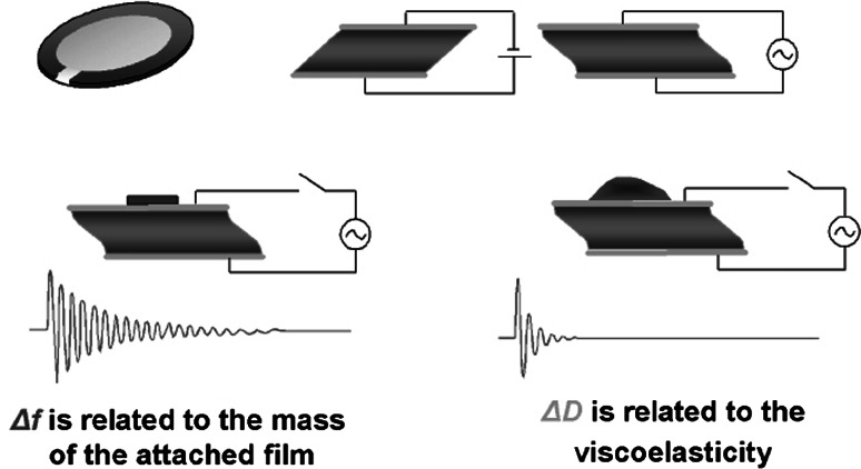
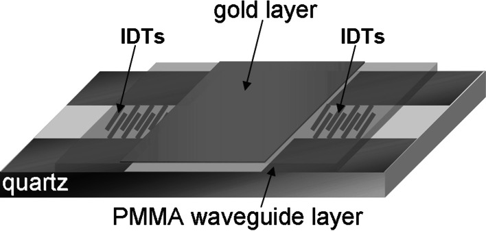
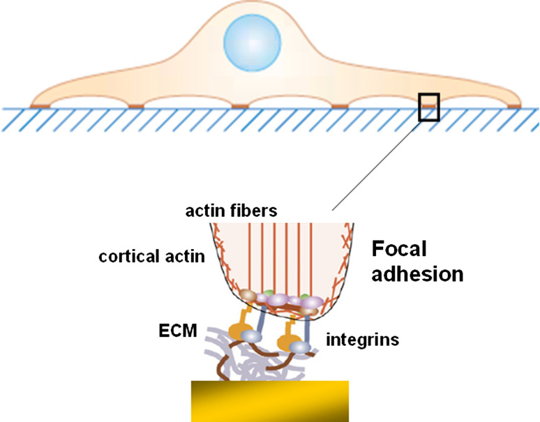
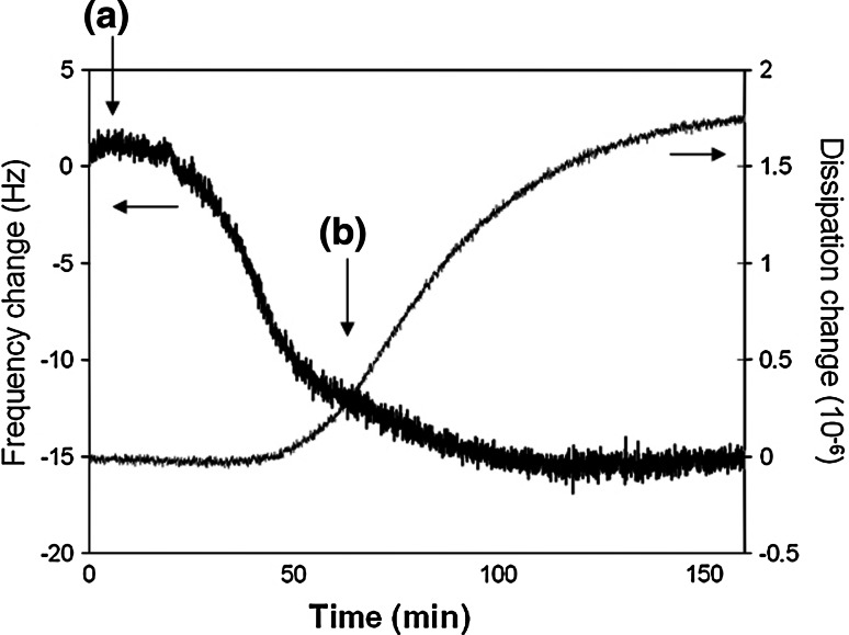
Acoustic biosensors offer the possibility to analyse cell attachment and spreading. This is due to the offered speed of detection, the real-time non-invasive approach and their high sensitivity not only to mass coupling, but also to viscoelastic changes occurring close to the sensor surface. Quartz crystal microbalance (QCM) and surface acoustic wave (Love-wave) systems have been used to monitor the adhesion of animal cells to various surfaces and record the behaviour of cell layers under various conditions. The sensors detect cells mostly via their sensitivity in viscoelasticity and mechanical properties. Particularly, the QCM sensor detects cytoskeletal rearrangements caused by specific drugs affecting either actin microfilaments or microtubules. The Love-wave sensor directly measures cell/substrate bonds via acoustic damping and provides 2D kinetic and affinity parameters. Other studies have applied the QCM sensor as a diagnostic tool for leukaemia and, potentially, for chemotherapeutic agents. Acoustic sensors have also been used in the evaluation of the cytocompatibility of artificial surfaces and, in general, they have the potential to become powerful tools for even more diverse cellular analysis.


Figures

Fig. 1
Fig. 1
Schematic illustration of the QCM-D principle of operation. The frequency of the oscillating crystal is related to the total oscillating mass, while the energy dissipation is related to the viscoelastic properties of the oscillating mass. Thus, changes in the mass induced upon adsorption of a rigid protein provide a change in frequency only, while if the mass is not rigid, which is often the case for biomolecules, there is a clear detectable change in the energy dissipation of the oscillating system. (Source: http://www.q-sense.com)
Fig. 2
Fig. 2
Schematic illustration of the acoustic Love-wave biosensor. The surface of the quartz-based acoustic-wave device, where two interdigitated transducers (IDTs) are deposited, is covered with a polymer guiding layer, in our case from poly-methylmethacrylate (PMMA). A thin layer of gold is deposited on top of the waveguide layer and between the IDTs. (Source: Saitakis et al. [41])
Fig. 3
Fig. 3
Focal adhesion of a cell on extracellular matrix proteins. Binding is mediated by integrins, which are intracellularly connected to the actin cytoskeleton
Fig. 4
Fig. 4
Real-time frequency and dissipation change of the QCM sensor during the addition of HLA-expressing human lymphoblastoid cells over an anti-HLA antibody-modified gold surface. a Addition of cell sample (106 cells ml−1), b wash with buffer
Fig. 5
Fig. 5
Schematic illustration of the sensor interface for cells interacting via membrane HLA-A2 molecules with surface-immobilised antibodies. Inside the cell, cortical actin microfilaments are shown. The two dashed lines indicate the penetration depths for the Love-wave (54 nm) and the QCM sensor (95 nm) (not drawn to scale). (Source: Saitakis et al. [41])
Fig. 6
Fig. 6
Time-dependent behavior of the Endothelial Cell QCM biosensor created by the addition of 2 × 104 endothelial cells (first arrowhead). The frequency (Δf) and resistance (ΔR) changes were recorded continuously until steady-state condition. Nocodazole was added to a final 2 μM concentration (second arrowhead). (Source: Marx et al. [59])
Fig. 7
Fig. 7
Amplitude change of the Love-wave sensor at equilibrium as a function of human lymphoblastoid cells added over the sensor surface (error bars correspond to standard deviation, n = 3 or 4) for three different numbers of the cell membrane HLA molecules. The values were: 3.7–5.7 × 105 HLA molecules per untreated cell, 1–10 × 104 molecules per mild acid-treated cell and 9.7–10.0 × 105 per cell from a high density culture. (Source: Saitakis et al. [49])

Similar articles

Cited by

References

    1. Gumbiner BM. Cell adhesion: the molecular basis of tissue architecture and morphogenesis. Cell. 1996;84(3):345–357. doi: 10.1016/S0092-8674(00)81279-9. - DOI - PubMed
    1. Alon R, Hammer DA, Springer TA. Lifetime of the P-selectin-carbohydrate bond and its response to tensile force in hydrodynamic flow. Nature. 1995;374(6522):539–542. doi: 10.1038/374539a0. - DOI - PubMed
    1. Berg EL, Goldstein LA, Jutila MA, Nakache M, Picker LJ, Streeter PR, Wu NW, Zhou D, Butcher EC. Homing receptors and vascular addressins: cell adhesion molecules that direct lymphocyte traffic. Immunol Rev. 1989;108:5–18. doi: 10.1111/j.1600-065X.1989.tb00010.x. - DOI - PubMed
    1. Chen S, Springer TA. Selectin receptor-ligand bonds: formation limited by shear rate and dissociation governed by the Bell model. Proc Natl Acad Sci USA. 2001;98(3):950–955. doi: 10.1073/pnas.98.3.950. - DOI - PMC - PubMed
    1. Juliano RL. Membrane receptors for extracellular matrix macromolecules: relationship to cell adhesion and tumor metastasis. Biochim Biophys Acta. 1987;907(3):261–278. - PubMed

Publication types

LinkOut - more resources