. 2011 Nov;22(11):2119-28.
doi: 10.1681/ASN.2011010069. Epub 2011 Oct 13.

APOL1 localization in normal kidney and nondiabetic kidney disease

Affiliations

APOL1 localization in normal kidney and nondiabetic kidney disease

Sethu M Madhavan et al. J Am Soc Nephrol. 2011 Nov.

Abstract

In patients of African ancestry, genetic variants in APOL1, which encodes apolipoprotein L1, associate with the nondiabetic kidney diseases, focal segmental glomerulosclerosis (FSGS), HIV-associated nephropathy (HIVAN), and hypertensive nephropathy. Understanding the renal localization of APOL1 may provide clues that will ultimately help elucidate the mechanisms by which APOL1 variants promote nephropathy. Here, we used immunohistology to examine APOL1 localization in normal human kidney sections and in biopsies demonstrating either FSGS (n = 8) or HIVAN (n = 2). Within normal glomeruli, APOL1 only localized to podocytes. Compared with normal glomeruli, fewer cells stained for APOL1 in FSGS and HIVAN glomeruli, even when expression of the podocyte markers GLEPP1 and synaptopodin appeared normal. APOL1 localized to proximal tubular epithelia in normal kidneys, FSGS, and HIVAN. We detected APOL1 in the arteriolar endothelium of normal and diseased kidney sections. Unexpectedly, in both FSGS and HIVAN but not normal kidneys, the media of medium artery and arterioles contained a subset of α-smooth muscle actin-positive cells that stained for APOL1. Comparing the renal distribution of APOL1 in nondiabetic kidney disease to normal kidney suggests that a previously unrecognized arteriopathy may contribute to disease pathogenesis in patients of African ancestry.


Figures
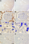
Figure 1.
Figure 1.
APOL1 localization in the normal adult human kidney using immunoperoxidase staining. Serial, formalin-fixed, paraffin-embedded sections of normal human kidney specimens were immunostained with rabbit polyclonal APOL1 antibody (A, C, E, & G) or purified rabbit polyclonal IgG as control (B, D, F, & H). (A) Low magnification image showing predominant APOL1 staining in the renal cortex, with relative sparing of medulla. (C) APOL1 expression is seen in a glomerulus and some cortical tubules. (E) Magnified image from boxed region in (C) shows APOL1 staining in a cell with anatomical position and morphologic characteristics of a podocyte, abutting the outer lamina of the glomerular basement membrane with red blood cells (asterisk) contained in a capillary loop. (G) APOL1 staining is also seen in extraglomerular arterial endothelium (arrow). Comparable images of normal kidney sections (B, D, F, & H) incubated with nonimmune purified rabbit polyclonal IgG have minimal staining consistent with residual endogenous peroxidase activity. Scale bars: 300 μm (A & B), 50 μm (C, D, G, & H).
Figure 2.
Figure 2.
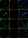
Colocalization of APOL1 with cell markers in the normal adult human kidney. Confocal immunofluorescent microscopy demonstrates (A, D, G, & J, green) APOL1 staining in the glomerulus of the normal kidney. (B & E, red) GLEPP1, a podocyte transmembrane protein tyrosine phosphatase, demarcates podocyte plasma membranes. (C & F) Merged images show that the APOL1 signal (green) is completely circumscribed by GLEPP1 staining (red) and appears to be present in the same cells. (D–F) Magnified view of the indicated region in (C) shows that APOL1 (green) localizes to two cell bodies (arrowheads) outlined by GLEPP1 staining (red), and nucleated endothelial cells lining the adjacent capillary loop (asterisks) show no APOL1 staining. (H) Cells within normal adult human glomeruli do not express α-SMA (red); however, positive α-SMA staining is seen in an adjacent arterial wall (double arrows). (I) Merged image shows APOL1 staining (green) in the adjacent arterial endothelium (arrow) but not within the arterial wall. (K & L, red) CD31 staining of the glomerular endothelium does not colocalize with APOL1 (green). The inset in (L) is a magnified image of the indicated region in that panel. (C, F, I, & L, blue) Nuclei were visualized with TOTO-3 staining. Scale bars: 50 μm (A–C & G–L), 5 μm (D–E).
Figure 3.
Figure 3.
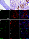
Altered renal localization of APOL1 in focal segmental glomerulosclerosis (FSGS). Immunoperoxidase (A–C) and confocal immunofluorescent images (D–L) of kidney biopsy specimens from patients with FSGS stained using anti-APOL1 antibody. (A) Reduced APOL1 staining is seen in glomeruli and cortical tubules compared with the normal kidney. In contrast, glomerular vascular tuft demonstrates prominent APOL1 expression (arrowheads). (B) Selected glomerulus from (A) shows diminished APOL1 staining (arrows); however, the juxtaglomerular arteriole wall is prominently APOL1 positive (arrowhead). (C) In contrast to the normal kidney, APOL1 also appears in the vessel wall of the renal arterioles. (D & F, green) APOL1 signal is diminished in the glomerulus, but glomerular GLEPP1 expression (E & F, red) is relatively preserved in early FSGS. The merged images (F) highlight these differences, where all APOL1 (green) positive cells stain for GLEPP1 (red) but not all GLEPP1 expressing cells stain for APOL1. (G–I) Residual glomerular APOL1 signal (G & I, green) in early FSGS does not colocalize with the α-SMA staining (H & I, red) of reactive mesangium. However, APOL1 signal (G & I, green) is observed in the wall of the arteriole at the vascular pole of the glomerulus (asterisk) and colocalizes with α-SMA (H & I, red), which labels vascular smooth muscle cells in the extraglomerular arteriolar wall (asterisk). (J–L, green) APOL1 remains diminished in early FSGS and does not colocalize with glomerular anti-CD31 staining (K & L, red), which identifies endothelial cells, but does colocalize in the extraglomerular vascular endothelium (arrowheads). (L) Merged image shows overlap of APOL1 (green) and CD31 (red) staining in the extraglomerular vascular endothelium (arrow) but not within the glomerulus. (F, I, & L, blue) Nuclei were visualized with TOTO-3 staining. Scale bars: 100 μm (A), 50 μm (B–L).
Figure 4.
Figure 4.
De novo localization of APOL1 to the renal arterial wall in FSGS. Confocal immunofluorescence imaging of medium sized renal arterioles from the normal human kidney (A–C) and from FSGS (D–F). (A & C, green) APOL1 signal is identified in cells anatomically consistent with endothelium of medium-sized renal arterioles (arrowheads) in the normal adult human kidney and adjacent tubular segments but does not colocalize with vascular smooth muscle cells of renal arterioles stained with anti-α-SMA antibody (B & C, red). APOL1-positive cells are located in the luminal wall of the blood vessel with surrounding α-SMA positive vascular smooth muscle cells, consistent with endothelial localization. (D & F, green) Representative cross-section of a renal arteriole from an FSGS biopsy demonstrates persistent endothelial APOL1 signal and de novo appearance within the vessel wall compared with the normal arteriole (A). (E & F, red) Anti-α-SMA antibody staining identifies APOL1-positive cells as vascular smooth muscle (F, green). The vascular endothelium remains positive for APOL1 staining (arrowhead). (C & F, blue) Nuclei were visualized with TOTO-3 staining. Scale bars: 50 μm (A–F).
Figure 5.
Figure 5.
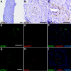
APOL1 redistribution is consistent between FSGS and HIVAN samples. Immunoperoxidase (A–C) and confocal immunofluorescent images (D–I) of kidney biopsy specimens from patients with HIVAN stained using anti-APOL1 antibody. (A) Reduced APOL1 staining in glomeruli and cortical tubules compared with the normal kidney. (B) Selected glomerulus from (A) shows diminished APOL1 staining (arrowhead). (C) In contrast to the normal kidney, APOL1 also appears in vessel wall of the renal arterioles. (D & F, green) APOL1 signal is diminished in the glomerulus, but remains colocalized with GLEPP1 expression (E & F, red), which is also diminished in HIVAN. (H & J, green) Glomerular APOL1 signal does not overlap with CD31 staining (I & J, red), which identifies glomerular capillary endothelium. (F & I, blue) Nuclei were visualized with TOTO-3 staining. Scale bars: 100 μm (A), 50 μm (B–I).
Figure 6.
Figure 6.
APOL1 is expressed in cultured human kidney cells and is induced in vascular endothelial cells with TNF/LPS treatment. (A) APOL1 is detectable by immunoblotting in cultured human podocytes, proximal tubule (HPRT) cells, and CHO cells transfected with an APOL1 expression construct. The blot is representative of three independent experiments. (B) APOL1 transcript is detectable in total RNA isolated from cultured human podocytes. The blot is representative of three independent experiments. (C, D) In vitro expression of APOL1 was detected in unstimulated and TNF- or LPS-stimulated human microvascular endothelial cells (hMVECs) by immunoblotting and normalized by the expression of tubulin (C). APOL1 expression was quantified by densitometry in unstimulated hMVECs and hMVECs stimulated with TNF or LPS (n = 3) (D). Results were normalized to tubulin expression and expressed as the mean ± SE; p values were calculated with ANOVA, followed by t test, to make intergroup comparisons. *P < 0.05.

Comment in

Similar articles

  • APOL1 genetic variants in focal segmental glomerulosclerosis and HIV-associated nephropathy.
    Kopp JB, Nelson GW, Sampath K, Johnson RC, Genovese G, An P, Friedman D, Briggs W, Dart R, Korbet S, Mokrzycki MH, Kimmel PL, Limou S, Ahuja TS, Berns JS, Fryc J, Simon EE, Smith MC, Trachtman H, Michel DM, Schelling JR, Vlahov D, Pollak M, Winkler CA. Kopp JB, et al. J Am Soc Nephrol. 2011 Nov;22(11):2129-37. doi: 10.1681/ASN.2011040388. Epub 2011 Oct 13. J Am Soc Nephrol. 2011. PMID: 21997394 Free PMC article.
  • Apolipoprotein l1 and the genetic basis for racial disparity in chronic kidney disease.
    Quaggin SE, George AL Jr. Quaggin SE, et al. J Am Soc Nephrol. 2011 Nov;22(11):1955-8. doi: 10.1681/ASN.2011090932. Epub 2011 Oct 13. J Am Soc Nephrol. 2011. PMID: 21997393 No abstract available.
  • APOL1 variants increase risk for FSGS and HIVAN but not IgA nephropathy.
    Papeta N, Kiryluk K, Patel A, Sterken R, Kacak N, Snyder HJ, Imus PH, Mhatre AN, Lawani AK, Julian BA, Wyatt RJ, Novak J, Wyatt CM, Ross MJ, Winston JA, Klotman ME, Cohen DJ, Appel GB, D'Agati VD, Klotman PE, Gharavi AG. Papeta N, et al. J Am Soc Nephrol. 2011 Nov;22(11):1991-6. doi: 10.1681/ASN.2011040434. Epub 2011 Oct 13. J Am Soc Nephrol. 2011. PMID: 21997397 Free PMC article.
  • APOL1 and kidney disease.
    Pollak MR, Genovese G, Friedman DJ. Pollak MR, et al. Curr Opin Nephrol Hypertens. 2012 Mar;21(2):179-82. doi: 10.1097/MNH.0b013e32835012ab. Curr Opin Nephrol Hypertens. 2012. PMID: 22257798 Review.
  • HIV-associated nephropathy: pathogenesis.
    Medapalli RK, He JC, Klotman PE. Medapalli RK, et al. Curr Opin Nephrol Hypertens. 2011 May;20(3):306-11. doi: 10.1097/MNH.0b013e328345359a. Curr Opin Nephrol Hypertens. 2011. PMID: 21358326 Free PMC article. Review.

Cited by

References

    1. Kao WH, Klag MJ, Meoni LA, Reich D, Berthier-Schaad Y, Li M, Coresh J, Patterson N, Tandon A, Powe NR, Fink NE, Sadler JH, Weir MR, Abboud HE, Adler SG, Divers J, Iyengar SK, Freedman BI, Kimmel PL, Knowler WC, Kohn OF, Kramp K, Leehey DJ, Nicholas SB, Pahl MV, Schelling JR, Sedor JR, Thornley-Brown D, Winkler CA, Smith MW, Parekh RS: MYH9 is associated with nondiabetic end-stage renal disease in African Americans. Nat Genet 40: 1185–1192, 2008 - PMC - PubMed
    1. Kopp JB, Smith MW, Nelson GW, Johnson RC, Freedman BI, Bowden DW, Oleksyk T, McKenzie LM, Kajiyama H, Ahuja TS, Berns JS, Briggs W, Cho ME, Dart RA, Kimmel PL, Korbet SM, Michel DM, Mokrzycki MH, Schelling JR, Simon E, Trachtman H, Vlahov D, Winkler CA: MYH9 is a major-effect risk gene for focal segmental glomerulosclerosis. Nat Genet 40: 1175–1184, 2008 - PMC - PubMed
    1. Arrondel C, Vodovar N, Knebelmann B, Grunfeld JP, Gubler MC, Antignac C, Heidet L: Expression of the nonmuscle myosin heavy chain IIA in the human kidney and screening for MYH9 mutations in Epstein and Fechtner syndromes. J Am Soc Nephrol 13: 65–74, 2002 - PubMed
    1. Ghiggeri GM, Caridi G, Magrini U, Sessa A, Savoia A, Seri M, Pecci A, Romagnoli R, Gangarossa S, Noris P, Sartore S, Necchi V, Ravazzolo R, Balduini CL: Genetics, clinical and pathological features of glomerulonephritis associated with mutations of nonmuscle myosin IIA (Fechtner syndrome). Am J Kidney Dis 41: 95–104, 2003 - PubMed
    1. Nelson GW, Freedman BI, Bowden DW, Langefeld CD, An P, Hicks PJ, Bostrom MA, Johnson RC, Kopp JB, Winkler CA: Dense mapping of MYH9 localizes the strongest kidney disease associations to the region of introns 13 to 15. Hum Mol Genet 19: 1805–1815, 2010 - PMC - PubMed

Publication types

MeSH terms

LinkOut - more resources