. 2012 Mar;46(3):323-30.
doi: 10.1165/rcmb.2011-0165OC. Epub 2011 Oct 13.

Hypoxic regulation of pulmonary vascular smooth muscle cyclic guanosine monophosphate-dependent kinase by the ubiquitin conjugating system

Affiliations

Hypoxic regulation of pulmonary vascular smooth muscle cyclic guanosine monophosphate-dependent kinase by the ubiquitin conjugating system

Ramaswamy Ramchandran et al. Am J Respir Cell Mol Biol. 2012 Mar.

Abstract

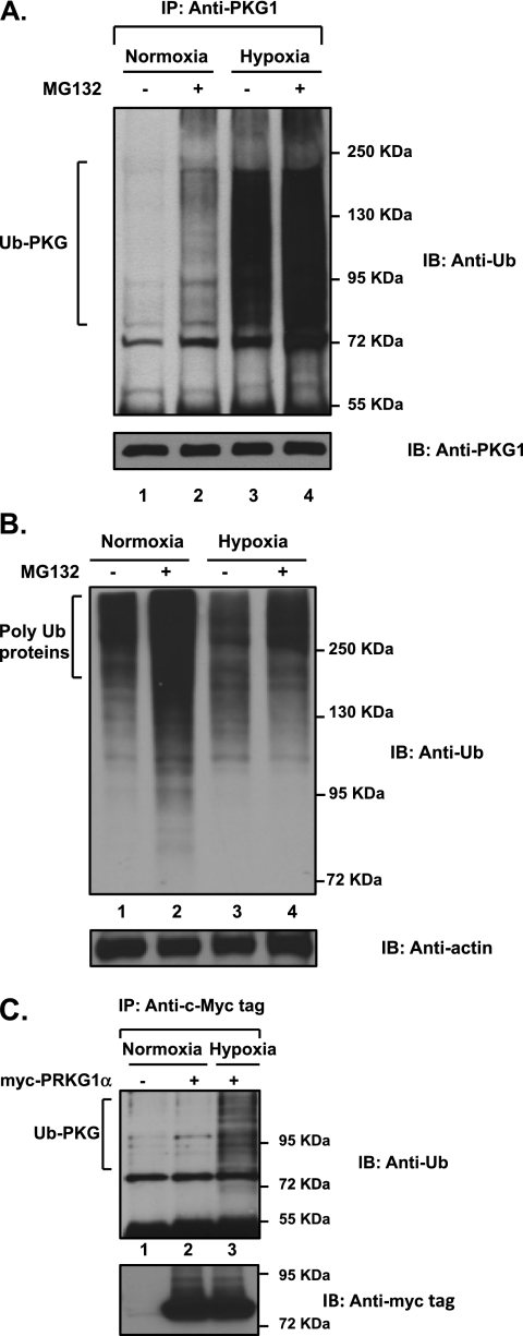
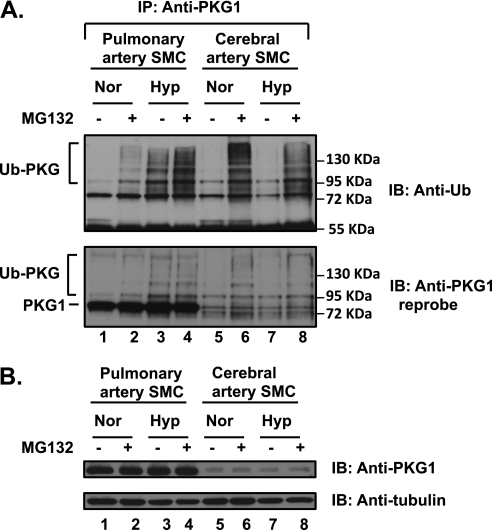
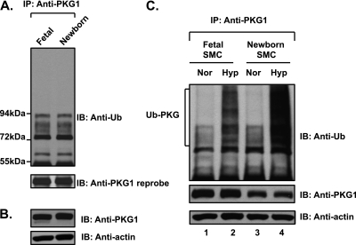
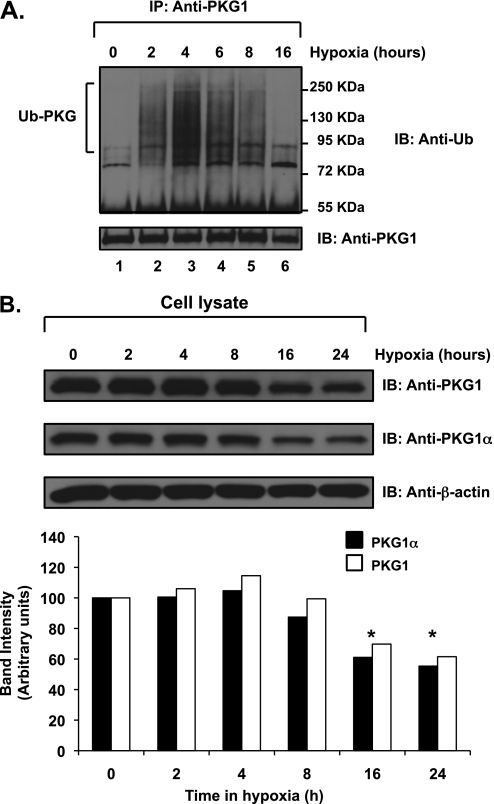
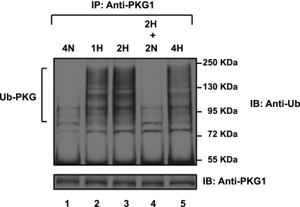
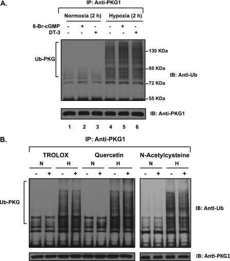
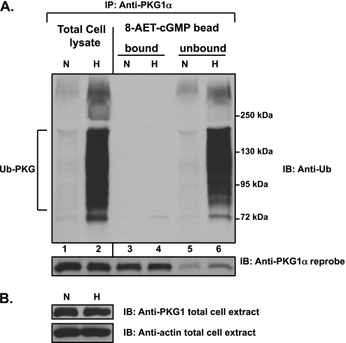
We previously reported that hypoxia attenuates nitric oxide-cyclic guanosine monophosphate (NO-cGMP)-mediated fetal pulmonary vessel relaxation by inhibiting cGMP-dependent protein kinase 1 (PKG1) activity, but not all the mechanisms by which acute hypoxia inhibits PKG1 activity have been delineated. Here we demonstrate for the first time, to the best of our knowledge, that acute hypoxia induces an accumulation of ubiquitinated PKG1 in ovine fetal and newborn pulmonary artery smooth muscle cells. Such a modification was not evident in ovine fetal systemic (cerebral) artery smooth muscle cells. The accumulation of polyubiquitinated PKG1 observed after 4 hours of hypoxia was affected neither by the activation of PKG1 kinase activity with the cell-permeable cGMP analogue 8-bromo-cGMP, nor by its inhibition with DT-3 in fetal pulmonary artery smooth muscle cells. Ubiquitinated PKG1α was unable to bind the cGMP analogue 8-(2-aminoethyl)thioguanosine-3',5' (AET)-cGMP, a ligand for the unmodified protein. Inhibition of the proteasomal complex with MG132 led to the accumulation of polyubiquitinated PKG1 in normoxia, indicating the involvement of the ubiquitin-26S proteasomal system in degradation and clearance of this protein under normoxic conditions. The ubiquitinated PKG1 under hypoxic conditions, however, was not predominantly targeted for proteasomal degradation. Importantly, reoxygenation reversed the acute hypoxia-induced accumulation of ubiquitinated PKG1. Our results suggest that the PKG1 ubiquitination induced by acute hypoxia plays a unique role in the regulation of the pulmonary vascular smooth muscle cell vasoreactivity and relaxation mediated by the NO-cGMP-PKG1 pathway.


Figures

Figure 1.
Figure 1.
Accumulation of the polyubiquitinated cyclic guanosine monophosphate (cGMP)–dependent protein kinase (PKG1) in acute hypoxia is not attributable to the inactivation of the proteasomal degradation pathway. Ovine fetal pulmonary artery smooth muscle cells were exposed to hypoxia or normoxia in the presence of the cell-permeable proteasome inhibitor MG132 (5 μM) for 4 hours. (A) Cell lysates were immunoprecipitated with a common PKG1 antibody and analyzed, using Western blotting with an anti-ubiquitin–specific monoclonal antibody. Blots were reprobed for concentrations of PKG1. Experiments were repeated three times, and representative blots are shown. An increased accumulation of ubiquitinated PKG1 (Ub-PKG) was observed after exposure to hypoxia. Treatment with MG132 indicates that PKG1 protein is targeted for proteasomal degradation under normoxic conditions, and an enhanced accumulation of Ub-PKG is observed during hypoxia. (B) Cell lysates were analyzed using Western blotting, and probed with anti-ubiquitin monoclonal antibody. (C) Exogenously transfected PKG1 is ubiquitin-modified during acute hypoxia in pulmonary artery smooth muscle cells. Fetal pulmonary artery smooth muscle cells (FPASMCs) were transiently transfected with the plasmid vector containing myc-PKG1α, as described in Materials and Methods, and exposed to normoxia or hypoxia for 4 hours. Cell extracts were immunoprecipitated with a c-Myc tag antibody and analyzed by Western blotting, using anti-ubiquitin antibody. Cell lysates containing the same amount of protein were probed with the c-Myc tag antibody. Experiments were repeated three times, and a representative blot is shown. IP, immunoprecipitation; IB, immunoblot.
Figure 2.
Figure 2.
Hypoxia-induced ubiquitination of PKG1 is specific to pulmonary artery smooth muscle cells (SMCs). Isolated FPASMCs and fetal cerebral artery smooth muscle cells (FCASMCs) were both exposed to hypoxia (Hyo) or normoxia (Nor) in the presence or absence of the proteasomal inhibitor MG132 (5 μM) for 4 hours. (A) The cell lysates were immunoprecipitated with the common PKG1 antibody and assessed using Western blotting with anti-ubiquitin monoclonal antibody, as described in Materials and Methods. Blots were stripped and reprobed with anti-PKG1 antibody. (B) The cell lysates were probed with a common PKG1 antibody, and tubulin served as the loading control. Experiments were repeated 3–4 times, and a representative blot is shown. The ubiquitination of PKG (Ub-PKG accumulation) was evident in hypoxia only in FPASMCs, and not in FCASMCs.
Figure 3.
Figure 3.
Fetal and newborn pulmonary arteries do not demonstrate increased ubiquitin modification of PKG1. The ubiquitin modification of PKG1 is increased in both fetal and newborn pulmonary artery SMCs upon exposure to acute hypoxia. (A) Fetal arteries were isolated under hypoxic conditions as in utero. Ovine fetal and newborn pulmonary artery extracts were immunoprecipitated with anti-PKG1 antibody and analyzed by Western blotting, using an anti-ubiquitin antibody. The blot was reprobed with anti-PKG1 antibody. (B) The arterial tissue extracts were analyzed by Western blotting, using anti-PKG1 and actin antibodies. (C) Fetal and newborn ovine pulmonary artery SMCs were exposed to hypoxia or normoxia for 3 hours. Cell lysates were immunoprecipitated with PKG1 antibody and assessed by Western blotting, using an anti-ubiquitin monoclonal antibody (top). Blots were reprobed with PKG1 antibody (middle). Actin in cell extracts served as loading control (bottom). A representative blot from three experiments is shown.
Figure 4.
Figure 4.
PKG1 is subject to time-dependant ubiquitination in acute hypoxia. Ovine fetal pulmonary vascular smooth muscle cells were exposed to normoxia or hypoxia. (A) Cell extracts were immunoprecipitated with a common PKG1 antibody and analyzed by SDS-PAGE and Western blotting, using an anti-ubiquitin monoclonal antibody. The blots were stripped and reprobed with PKG1 antibody. (B) Cell lysates were analyzed by Western blotting, using a common PKG1 antibody or PKG1α isoform–specific antibody. Gel loading controls were assessed by probing with an actin antibody. Bottom: A plot of the relative PKG1 protein band intensities, normalized to actin levels, was quantified using ImageJ software (National Institutes of Health), and expressed as a percentage. Experiments were repeated 4–5 times, and representative blots are shown. *P < 0.05. The data shown are for hypoxia experiments. No PKG1 ubiquitination during normoxia was observed (data not shown).
Figure 5.
Figure 5.
Reoxygenation reverses acute hypoxia-induced PKG1 ubiquitin modification. FPASMCs were exposed to hypoxia for 2 hours and transferred to normoxia for an additional 2 hours. Cell lysates were immunoprecipitated with PKG1 antibody and analyzed according to Western blotting, using anti-ubiquitin monoclonal antibody, and were reprobed with PKG1 antibody. The experiment was repeated three times, and a representative blot is shown. Duration of treatment time in hours is indicated. H, hypoxia (3% O2); N, normoxia (21% O2). Hypoxic cells that were reoxygenated contained no ubiquitinated PKG1 (Ub-PKG), similar to normoxic cells that had never been exposed to hypoxia.
Figure 6.
Figure 6.
Hypoxia-induced ubiquitin modification of PKG1 is unaffected by the endogenous activation or inhibition of its kinase activity or by common scavengers of reactive oxygen species or peroxynitrite. (A) FPASMCs were exposed to hypoxia for 2 hours, with or without the cell-permeable PKG1 inhibitor DT-3 (5 μM), or the phosphodiesterase-resistant activator 8-bromo (Br)-cGMP (0.5 mM). The cell lysates were immunoprecipitated with PKG1 and assessed by Western blotting, using an anti-ubiquitin monoclonal antibody. The blots were also reprobed with PKG1 antibody. The level of PKG activity exerted no effect on the accumulation of ubiquitinated PKG1 (Ub-PKG) during acute hypoxia. (B) FPASMCs were exposed to hypoxia or normoxia for 3 hours, with or without 0.2 mM trolox, 0.1 mM quercetin, or 10 mM N-acetylcysteine. Cells were preincubated for 30 minutes in normoxia (21% oxygen and 5% CO2) with the different scavengers before exposure to hypoxia (3% O2 and 5% CO2) in the presence of the same scavenger. The cell lysates were immunoprecipitated with PKG1 and assessed by Western blotting, using an anti-ubiquitin monoclonal antibody. The blots were also reprobed with PKG1 antibody. A representative blot from 3–4 experiments is shown.
Figure 7.
Figure 7.
Hypoxia-induced PKG1 ubiquitination hinders its binding to 8-(2-aminoethyl)thioguanosine-3′,5′ (AET)-cGMP. Cell extracts were prepared from FPASMCs exposed to hypoxia (3% O2, 3 hours) in buffer A, as described in Materials and Methods. (A) The extracts were incubated with 8-AET-cGMP agarose beads. After washing the beads, the bound proteins were eluted with 25 mM cGMP. The total cell lysates, namely, the bead “bound” and “unbound” proteins, were immunoprecipitated with PKG1α antibody and assessed by Western blotting, using an anti-ubiquitin antibody. The blots were stripped and reprobed with PKG1α antibody. (B) The cell extracts were probed for PKG1 and actin. All experiments were repeated 3–4 times. A representative blot is shown. Ubiquitinated PKG1α (Ub-PKG) from hypoxically exposed cells exhibited defective binding to 8-AET-cGMP, compared with native, unmodified PKG1α.

References

    1. Hofmann F, Bernhard D, Lukowski R, Weinmeister P. cGMP regulated protein kinases (CGK). Handb Exp Pharmacol 2009;191:137–162 - PubMed
    1. Schlossmann J, Desch M. CGK substrates. Handb Exp Pharmacol 2009;191:163–193 - PubMed
    1. Francis SH, Busch JL, Corbin JD, Sibley D. cGMP-dependent protein kinases and cGMP phosphodiesterases in nitric oxide and cGMP action. Pharmacol Rev 2010;62:525–563 - PMC - PubMed
    1. Hofmann F, Feil R, Kleppisch T, Schlossmann J. Function of cGMP-dependent protein kinases as revealed by gene deletion. Physiol Rev 2006;86:1–23 - PubMed
    1. Surks HK, Mochizuki N, Kasai Y, Georgescu SP, Tang KM, Ito M, Lincoln TM, Mendelsohn ME. Regulation of myosin phosphatase by a specific interaction with cGMP-dependent protein kinase Ialpha. Science 1999;286:1583–1587 - PubMed

Publication types

MeSH terms