Review
. 2013 Feb;62(2):310-6.
doi: 10.1136/gutjnl-2011-301008. Epub 2011 Oct 13.

Oncogenic transcription factors: cornerstones of inflammation-linked pancreatic carcinogenesis

Affiliations
Review

Oncogenic transcription factors: cornerstones of inflammation-linked pancreatic carcinogenesis

Sandra Baumgart et al. Gut. 2013 Feb.

Abstract

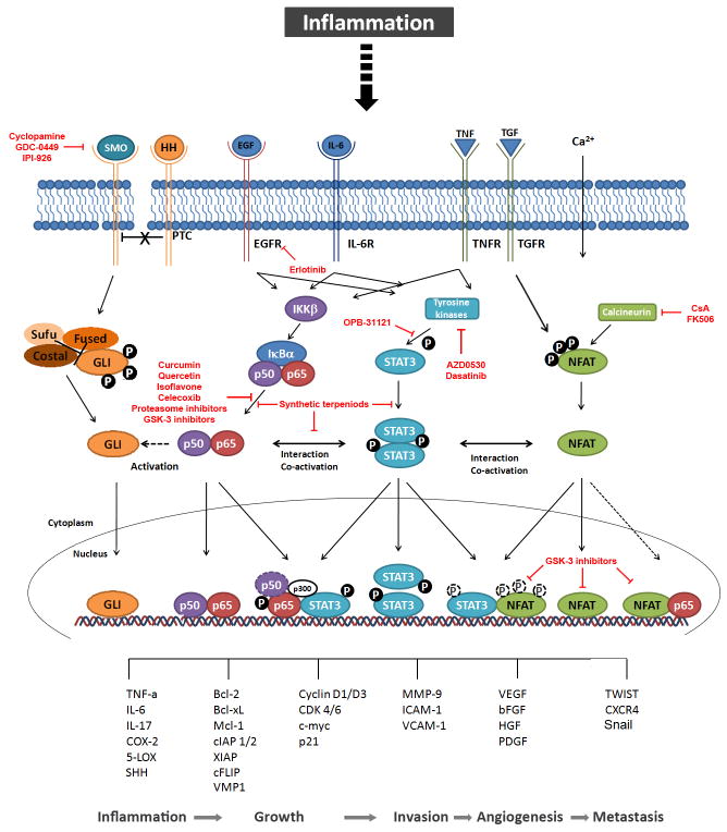
Transcription factors are proteins that regulate gene expression by modulating the synthesis of messenger RNA. Since this process is often one dominant control point in the production of many proteins, transcription factors represent the key regulators of numerous cellular functions, including proliferation, differentiation and apoptosis. Pancreatic cancer progression is characterised by activation of inflammatory signalling pathways converging on a limited set of transcription factors that fine-tune gene expression patterns contributing to the growth and maintenance of these tumours. Thus strategies targeting these transcriptional networks activated in pancreatic cancer cells could block the effects of upstream inflammatory responses participating in pancreatic tumorigenesis. The authors review this field of research and summarise current strategies for targeting oncogenic transcription factors and their activating signalling networks in the treatment of pancreatic cancer.


Figures

Figure 1
Figure 1. Putative crosstalk between oncogenic transcription factors contributing to pancreatic carcinogenesis
Inflammation induces binding of cytokines and inflammatory ligands to their receptors, which in turn activate the corresponding transcription factor signaling pathways. Binding of HH to the transmembrane protein PTC reverses the inhibitory effect on SMO, which again releases GLI transcription factors from their inhibitory complex and finally initiates nuclear translocation. The activation of the pathway can be blocked by inhibitors of SMO such as cyclopamine and its derivatives currently in clinical studies, GDC-0449 and IPI-926. Interactions with the NF-κB signaling pathway are illustrated by the dashed arrowhead which indicates activation of the HH pathway mainly through the regulation of the expression of its ligand SHH. Binding of EGF or IL-6 activates IKKβ kinase which stimulates degradation of IκBα. Subsequently, NF-κB proteins are released to the nucleus, where they mediate gene transcription alone or in cooperation with STAT proteins. Curcumin, Quercetin, Isoflavone and proteasome or GSK-3 inhibitors interfere with NF-κB signaling mainly through inhibition of NF-κB activation. Thereby, p-STAT3 binding to p-p65 induces histone acetyl transcerase p300 and retains active p65 in the nucleus. Synthetic terpenoids can either block isolated NF-κB and STAT3 activation, or inhibit the interaction of both pathways. A putative binding of p50 to STAT3 is shown by the dashed circle. The STAT3 signaling pathway can as well be activated by the aforementioned inflammatory stimuli. Upon binding of the ligands to the transmembrane receptors (receptor-bound) kinases, such as JAKs, SRC or ABL, are activated and in turn phosphorylate STAT3 proteins. EGFR inhibitor erlotinib as well as tyrosine kinase inhibitors AZD0530 and dasatinib have shown to efficiently block STAT3 activation. The subsequent dimerization leads to nuclear translocation and target gene transcription, partially by interaction with NFAT transcription factors. The interaction on target promoters is not dependent on STAT and NFAT phosphorylation, illustrated by dashed circles. The canonical NFAT signaling pathway is activated by intracellular Ca2+ rises leading to activation of the phosphatase Calcineurin and dephosphorylation of NFAT proteins which shuttle to the nucleus and bind to their target promoters. Calcineurin inhibitors CsA and FK506 block NFAT dephosphorylation and nuclear translocation. NFAT phosphorylation does not exclude nuclear localization and binding to partner proteins, shown by dashed white circles. The dashed arrow indicates a possible interaction with p65, which has been demonstrated for other non-pancreatic tissues. GSK-3 inhibitors successfully disrupt NFAT binding to partner proteins such as STAT3 as well as NFAT transcriptional activity in the nucleus. Finally, the network of inflammatory transcription factors regulate numerous target genes mediating inflammation, growth, invasion, angiogenesis and metastasis, thereby contributing to pancreatic carcinogenesis. PTC, indicates Patched; SMO, Smoothened; HH, Hedgehog; Sufu, SHH, Sonic HH; Suppressor of Fused; EGF(R), epidermal growth factor receptor; IL-6(R), interleukin-6 (receptor); IKKβ, IκB kinase β; IκBα, inhibitor of NF-κB α; GSK-3, Glycogen synthase kinase-3; STAT3, signal transducer and activator of transcription 3; JAK, janus kinase; TNF(R), tumor necrosis factor (receptor); TGF(R), transforming growth factor (receptor); NFAT, nuclear factor of activated T-cells; CsA, Cyclosporine A; COX-2, cyclooxygenase-2; 5-LOX, 5-lipooxygenase; Bcl-2/xL, B cell lymphoma 2/xL; Mcl-1, myeloid cell leukemia sequence 1; cIAP 1/2, cellular inhibitor of apoptosis; XIAP, X-linked inhibitor of apoptosis protein; cFLIP, Cellular FLICE (FADD-like IL-1beta-converting enzyme)-inhibitory protein; CDK 4/6, cyclin-dependent kinase 4/6; MMP-9, matrix-metaloproteinase-9; ICAM-1, inter-Cellular Adhesion Molecule 1;VCAM-1, vascular cell adhesion molecule 1; VEGF, vascular endothelial growth factor; bFGF, basic fibroblast growth factor; HGF, hepatocyte growth factor; VMP1, Vacuole Membrane Protein 1; PDGF, platelet-derived growth factor; CXCR4, CXC-motif chemokine receptor 4.

References

    1. Löhr M, Klöppel G, Maisonneuve P, et al. Frequency of K-ras mutations in pancreatic intraductal neoplasias associated with pancreatic ductal adenocarcinoma and chronic pancreatitis: a meta-analysis. Neoplasia. 2005;7:17–23. - PMC - PubMed
    1. Hingorani SR, Petricoin EF, Maitra A, et al. Preinvasive and invasive ductal pancreatic cancer and its early detection in the mouse. Cancer Cell. 2003;4:437–450. - PubMed
    1. Morris JP, 4th, Wang SC, Hebrok M. KRAS, Hedgehog, Wnt and the twisted developmental biology of pancreatic ductal adenocarcinoma. Nat Rev Cancer. 2010;10:683–695. - PMC - PubMed
    1. Guerra C, Schuhmacher AJ, Cañamero M, et al. Chronic pancreatitis is essential for induction of pancreatic ductal adenocarcinoma by K-Ras oncogenes in adult mice. Cancer Cell. 2007;11:291–302. - PubMed
    1. Erkan M, Reiser-Erkan C, Michalski CW, et al. Tumor microenvironment and progression of pancreatic cancer. Exp Oncol. 2010;32:128–131. - PubMed

Publication types

MeSH terms

Substances