

"Stealth" melanoma cells in histology-negative sentinel lymph nodes
- PMID: 21997686
- PMCID: PMC3203732
- DOI: 10.1097/PAS.0b013e3182322cf7
"Stealth" melanoma cells in histology-negative sentinel lymph nodes
Abstract
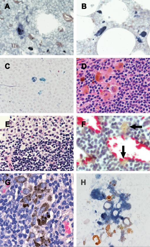
A proportion of patients who develop regional and distant recurrences of melanoma after a pathologically negative sentinel lymph node (SN) biopsy are reported to have enhanced signals for melanoma-associated messenger ribonucleic acid (mRNA) when sensitive molecular approaches such as reverse transcriptase polymerase chain reaction (RT-PCR) are used to evaluate their SN tissue. The significance of these findings remains controversial, because the cellular source of the augmented signals cannot be known as the nodal tissue is destroyed during preparation for RT-PCR. Nevertheless, it is claimed that the source of the augmented signal is covert metastatic melanoma cells. To determine whether there are histologically occult metastases in SN and whether there are sources of augmentable melanoma-associated mRNA other than melanoma cells, we applied reverse transcriptase in situ polymerase chain reaction (RT in situ PCR) to formalin-fixed paraffin-embedded nodal tissue. This approach amplifies small amounts of melanoma-associated mRNA and permits identification of cells that express that mRNA. Cells containing MART-1 mRNA were detected in 6 of 21 SNs (29%) and 2 of 16 nonsentinel lymph node (NSNs) (13%) that were tumor negative on hematoxylin and eosin and on immunohistochemical assessment for S-100, MART-1, and HMB-45. In patients with microscopic evidence of melanoma in their SN, MART-1 mRNA-positive cells were identified in 2 of 7 NSNs (29%) that were histologically tumor free. MART-1 mRNA-positive cells were also detected in tumor-negative SN sections from 6 of 7 (86%) nodes that had tumor present in areas of the node not represented in the studied sections. Some cells that expressed MART-1 mRNA that was diffusely distributed in the cytoplasm appeared to be melanoma cells, whereas others resembled macrophages. The latter cells expressed augmented mRNA on granules that were intermixed with melanin granules. In other cases, MART-1 mRNA-positive macrophage-like cells contained nuclei and nucleoli more typical of melanoma cells and may represent the macrophage-melanoma hybrids that have been previously reported. Combination of RT in situ PCR for MART-1 mRNA and immunohistochemistry for CD68 revealed that CD68 was colocalized in some cells that expressed MART-1 mRNA. Some lymph nodes that are tumor negative by histology and immunohistochemistry contain cells that express mRNA for MART-1. Some of these cells may be interpreted as "stealth" melanoma cells in which, despite the presence of MART-1 mRNA, there is an absence of immunohistochemically detectable MART-1 protein. Other cells that contain MART-1 mRNA are clearly not melanoma cells or may represent melanoma hybrids. These findings should be taken into account when interpreting and applying the results of RT-PCR analysis of nodal (and other) tissues.
Figures

Similar articles
-
Multimarker reverse transcriptase-polymerase chain reaction assay in lymphatic drainage and sentinel node tumor burden.Ann Surg Oncol. 2010 Dec;17(12):3314-23. doi: 10.1245/s10434-010-1142-9. Epub 2010 Jul 7. Ann Surg Oncol. 2010. PMID: 20607422 Free PMC article.
-
Pathologic assessment of melanoma sentinel nodes: a role for molecular analysis using quantitative real-time reverse transcription-PCR for MART-1 and tyrosinase messenger RNA.Clin Cancer Res. 2005 Feb 15;11(4):1425-33. doi: 10.1158/1078-0432.CCR-04-1193. Clin Cancer Res. 2005. PMID: 15746042
-
Evaluation of micrometastases in sentinel lymph nodes of cutaneous melanoma: higher diagnostic accuracy with Melan-A and MART-1 compared with S-100 protein and HMB-45.Am J Surg Pathol. 2001 Aug;25(8):1039-46. doi: 10.1097/00000478-200108000-00008. Am J Surg Pathol. 2001. PMID: 11474288
-
Pathologic examination of sentinel lymph nodes from melanoma patients.Semin Diagn Pathol. 2008 May;25(2):100-11. doi: 10.1053/j.semdp.2008.04.002. Semin Diagn Pathol. 2008. PMID: 18697713 Review.
-
Detection of micrometastasis in sentinel lymph nodes of patients with primary cutaneous melanoma.Recent Results Cancer Res. 2001;158:137-46. doi: 10.1007/978-3-642-59537-0_14. Recent Results Cancer Res. 2001. PMID: 11092041 Review.
Cited by
-
Missing sentinel lymph node in cutaneous melanoma.Indian J Nucl Med. 2015 Apr-Jun;30(2):158-61. doi: 10.4103/0972-3919.152982. Indian J Nucl Med. 2015. PMID: 25829738 Free PMC article.
-
Analysis of melanoma recurrence following a negative sentinel lymph node biopsy.Melanoma Manag. 2015 Aug;2(3):285-294. doi: 10.2217/mmt.15.19. Epub 2015 Aug 10. Melanoma Manag. 2015. PMID: 30190855 Free PMC article. Review.
-
Tumor-Cell-Macrophage Fusion Cells as Liquid Biomarkers and Tumor Enhancers in Cancer.Int J Mol Sci. 2020 Mar 9;21(5):1872. doi: 10.3390/ijms21051872. Int J Mol Sci. 2020. PMID: 32182935 Free PMC article. Review.
-
A Melanoma Brain Metastasis with a Donor-Patient Hybrid Genome following Bone Marrow Transplantation: First Evidence for Fusion in Human Cancer.PLoS One. 2013 Jun 26;8(6):e66731. doi: 10.1371/journal.pone.0066731. Print 2013. PLoS One. 2013. PMID: 23840523 Free PMC article.
-
Melanoma-Derived BRAF(V600E) Mutation in Peritumoral Stromal Cells: Implications for in Vivo Cell Fusion.Int J Mol Sci. 2016 Jun 21;17(6):980. doi: 10.3390/ijms17060980. Int J Mol Sci. 2016. PMID: 27338362 Free PMC article.
References
-
- Abdelatif OM, Khankhanian NK, Crosby JH, et al. Malignant fibrous histiocytoma and malignant melanoma: the role of immunohistochemistry and electron microscopy in the differential diagnosis. Mod Pathol. 1989;2:477–485. - PubMed
-
- Adam PJ, Boyd R, Tyson KL, et al. Comprehensive proteomic analysis of breast cancer cell membranes reveals unique proteins with potential roles in clinical cancer. J Biol Chem. 2003;278:6482–6489. - PubMed
-
- Blaheta HJ, Ellwanger U, Schittek B, et al. Examination of regional lymph nodes by sentinel node biopsy and molecular analysis provides new staging facilities in primary cutaneous melanoma. J Invest Dermatol. 2000;114:637–642. - PubMed
-
- Bonin S, Niccolini B, Calacione R, et al. Molecular analyses of sentinel lymph nodes: an open question. J Eur Acad Dermatol Venereol. 2002;16:34–39. - PubMed
-
- Bostick PJ, Morton DL, Turner RR, et al. Prognostic significance of occult metastases detected by sentinel lymphadenectomy and reverse transcriptase-polymerase chain reaction in early-stage melanoma patients. J Clin Oncol. 1999;17:3238–3244. - PubMed
Publication types
MeSH terms
Substances
Grants and funding
LinkOut - more resources
Full Text Sources
Other Literature Sources
Medical
